Vanillin Assay-Modified Protocol for Longitudinal Nutrition Utilization Study
Vanessa Corby-Harris
Fat Body
Grand Challenge
Honey Bee
Lipid
Folch
Vanillin
Tissue
NP305
2022-21000-022-000D
Vanillin Assay
Crop Production
Abstract
This protocol is a modified version of the vanillin assay based off Van Handel's 1985 manuscript: "Rapid Determination of Total Lipids in Mosquitoes."
This method is tailored for determining total lipids in Honey Bee tissues. Here, dry weight material is reacted with vanillin sulfuric acid to form a colorimetric change proportional to the amount of lipid in the material. It is then compared against a known (lipid) external standard to give the total lipid in µg/µL of material.
Before start
One plate can run 23 samples. Do not process more than 23 samples at a time. It takes ~3 .5 hours from
beginning with bead-beaten samples to step #18. It takes ~2 hours to finish the assay after
drying
Steps
Tissue Sample Prep
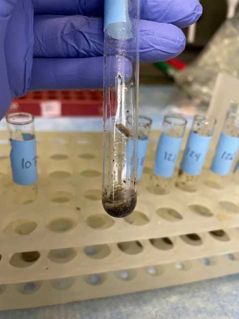
Label a 2 ml bead beating tube then add 3 x 3.2mm chrome steel beads into each vial.
Weigh the tube and record the weight.
Place tissue into pre-weighed bead beating tube.


Place tissue samples in drying oven for 96 hours at 60 degrees C. When dry weigh the tubes and record the dry mass.
Vanillin Reagent Preparation
Prepare 2:1 chloroform:methanol
■ 200 mL chloroform
■ 100 mL methanol
Prepare 0.25% KCl
■ 0.25 g KCl
■ 100 mL distilled water
Prepare vanillin-phosphoric acid
○ Use an amber bottle or wrap a clear bottle in foil
○ Dissolve 600 mg vanillin in 100 mL hot water
○ Add 400 mL 85% phosphoric acid
○ Store in the dark. Reagent is stable for several months, but should be discarded once it darkens
Standard Curve Preparation
Prepare standards using soybean oil (i.e. Wesson or Crisco) dissolved in chloroform (use 1 mL tip for chloroform). Soybean oil is ~747 g/L of unsaturated fatty acids double check the value for whatever brand you use.
To make 7000ul of a 7.5ug/ul solution of lipid in chloroform, add 70ul of soybean oil to 6930ul of chloroform.
Now, make serial dilutions:
o Make a 3.75ug/ul concentration (½ dilution of the 7.5ug/ul) by adding 1 part of the 7.5ug/ul (strongest standard) to 1 part chloroform.
o Make a 1.875ug/ul concentration (½ dilution of the 3.75ug/ul) by adding 1 part of the 3.75ug/ul to 1 part chloroform.
o Make a 0.9375ug/ul concentration (½ dilution of the 1.875ug/ul) by adding 1 part of the 1.875ug/ul to 1 part chloroform.
o Make a 0.46875ug/ul concentration (½ dilution of the 0.9375ug/ul) by adding 1 part of the 0.9375ug/ul to 1 part chloroform.
o Make a 0.234ug/ul concentration (½ dilution of the 0.46875ug/ul) by adding 1 part of the 0.46875ug/ul to 1 part chloroform.
o Make a 0.117ug/ul concentration (½ dilution of the 0.234ug/ul) by adding 1 part of the 0.234ug/ul to 1 part chloroform.
Add 100 uL of each of the 7 standards to empty screw cap vials.
o Adding 100ul of 3.75ug/ul = 3.75ug/ul x 100ul = 375ug of lipid in that well.
o Adding 100ul of 1.875ug/ul = 1.875ug/ul x 100ul = 187.5ug of lipid in that well.
o Etc…
Vanillin Protocol for Tissues
Pour entire contents of bead beating tube (see description) to a new glass culture tube (16x100mm). Add 1ml water to original vial and wash to get all contents out of the tube. Repeat 4x.
Add 1000ul 2:1 chloroform:methanol to each tube.
Add 210ul 0.25% KCl to each culture tube sample and vortex for ≥5 seconds.
Centrifuge these samples at 2000 rpm for 15 minutes. Move the chloroform layer (the bottom layer) to a labeled glass screw cap vial.
Repeat steps 13-15 for a total of 2 washes. Go through the vials and remove any residual top layer that accidentally got moved over with the chloroform layer.
Vanillin Protocol Continued
Add 100 uL of just chloroform to another empty screw cap vial. This is the negative control.
Dry the samples from step 16, one of each standard, and the negative control to completion. This should take roughly 1.5-2 hours depending on the number of samples if using a speed vac.
Alternatively drying can be achieved by leaving the samples in the fume hood overnight with the lids off but may require additional vacuuming the next morning.
Important: Make sure the speed vac is evenly balanced so as to not break the rotor! Do not use heat!
After the samples are dried, turn on the heating block to 100°C.
Add 182 ul of concentrated sulfuric acid to each vial and place the screw cap back on. Caution: sulfuric acid is very caustic!
Heat the samples at 100°C for 15 minutes. The standards and the samples should start to develop a brown color.
Take the samples off of heating block. Turn off and unplug the heating block once you are done using it.
Add 1480 ul of vanillin-phosphoric acid to each vial. Cap each vial and briefly vortex on low to mix the sample thoroughly.
Cover the rack and vials with foil and leave at room temperature for 15 mins.
If the samples and standards are very dark and highly concentrated, they need to be diluted to be within range of the spectrophotometer. If needed, add 100uL of each sample with sulfuric acid and lipids to appropriate amount of additional vanillin-phosphoric acid in a centrifuge tube. We typically need to dilute 10 bee abdominal carcasses by 8 to be within the range of the assay.
Make your plate map.
Add 100 ul of each sample in triplicate to the plate wells.
Read the plate at 525 nm and save the data.
Map your standard curve (Y axis=absorbance, X axis = ug/ul). Use equation for linear curve to infer the ug/ul values for your samples from the standard curve. This value (a concentration) should be normalized for the dry weight of tissue used in the reaction to yield the amount of lipid per unit of tissue. A good reference for how to do this is Vaudo et al. (2020) "Pollen protein:lipid macronutrient ratios may guide broad patterns of bee species floral preferences." Insects. 11 (2), 132, (2020).
