Immunofluorescence Staining
Hemanth Ramesh Nelvagal, Toby J Curless, Zane Jaunmuktane
Abstract
The protocol describes immunofluorescence staining on brain sections.
Steps
Preparation
Generate tissue sections using standard microtome sectioning protocols.
Heat tissue dry tissue sections for 1h 0m 0s at 60°C
De-paraffinisation and Immunofluorescence
De-paraffinise sections by 0h 5m 0s min washes in xylene (x3), 0h 5m 0s 100 % ethanol (x1), 0h 5m 0s 70 % ethanol (x1) and 0h 5m 0s DEPC H2O (x1).
Rinse sections in PBS.
Encircle tissue with a hydrophobic ImmEdgeTM PAP pen (Vector laboratories) to contain solution.
Incubate tissue with H2O2for 0h 15m 0s mins at room temperature.
Wash tissue 3x in PBS.
Antigen retrieval – heat 1 container (250 ml) DEPC H2O, and 1 container (250 ml) CC1 buffer to
99°C using a steamer (BRAUN tribute collection).
Add slides to heated DEPC H2O for 10 s, then transfer to CC1 buffer and incubate at 99°C for 0h 15m 0s.
Remove slides and was 3x with PBS for 0h 5m 0s mins total.
Block sections with 10 % normal goat serum in PBS for 2h 0m 0s.
Wash tissue in PBS.
Incubate sections in primary antibodies (antibodies diluted in PBS) at 4°C 2h 0m 0s.
Following overnight incubation, wash the sections in PBS 3x for a total of0h 15m 0s and shake off excess PBS after each 0h 5m 0s interval.
Incubate sections with 1:100 secondary antibodies (e.g., Alexa Fluor 488-conjugated anti-mouse secondary antibody (Invitrogen) and Alexa Fluor 568-conjugated anti-rabbit secondary antibody (Invitrogen)) (for 0h 30m 0s mins at room temperature in the dark.
Wash sections in PBS 3x for a total of 0h 15m 0s – whilst incubating, remove Antifade mountant (P36980, Thermo Fisher Scientific) from refrigerator and bring to room temperature.
Counterstain sections with Hoechst (1:15000 (diluted in DEPC H2O)) for 0h 0m 30s sec
4°C Rinse sections in PBS 2x for a total of 0h 6m 0s
Coverslip slides using Antifade mountant (P36980, Thermo Fisher Scientific) and store slides in the dark at 4°C.
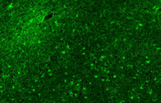
Capture slides on an epifluorescence microscope (Leica) using Alexa 488 filters.