TNF ELISA Protocol
Caitlyn Henry
Abstract
The purpose of this experiment is to investigate the effects of forskolin-mediated cAMP activation on TNF-ɑ secretion by LPS-treated Schwann cells. The immortalized rat RT4-D6P2T cell line (ATCC #CRL-2768) was cultured and received one of the following treatments: 0.1, 1, or 10 μg/mL of LPS, in N2 media (control) or N2 media supplemented with 2 μM of forskolin, for 3 hours. Cell media samples were collected, and the Rat TNF-alpha ELISA kit (RayBiotech, Cat #ELR-TNFa-1, Norcross, GA) was performed to quantify changes in TNF-ɑ secretion in response to the different treatment combinations.
Steps
To prepare RT4-D6P2T cell media samples (for three 6-well plates):
Aseptically culture immortalized rat RT4-D6P2T Schwann cells (ATCC, Cat #CRL-2768, Manassas, VA) in Dulbecco's Modified Eagle Medium (DMEM) (ATCC, Cat #30-2002, Manassas, VA) supplemented with 10% fetal bovine serum (FBS) (Thermo Fisher, Cat #16000044, Waltham, VA) and 1% penicillin/streptomycin (Pen-strep) (GIBCO, Cat #15140-015, Gaithersburg, MD)/amphotericin B (R&D Systems, Cat #B23192, Minneapolis, MN) at 37°C and 5% CO2 in poly-L-lysine (PLL)-coated dishes.
At 80% confluency, split and seed cells into DMEM (2 mL DMEM/well) in three PLL-coated 6-well plates at a density of ~300,000 cells/well.
Incubate cells in DMEM for 24 hours.
After 24 hours, aspirate the DMEM and wash each well 2-3x with 2 mL HBSS. After the last wash, add 2 mL N2 media (DMEM/F12, no phenol red [Thermo Fisher, Cat #21041025, Waltham, MA] supplemented with 5 μg/mL insulin [Sigma, Cat #91077C, St. Louis, MO] and 100 μg/mL apo-transferrin [Sigma, Cat #T1147, St. Louis, MO]) to each well.
Incubate cells in N2 media for 24 hours.
After 24 hours, prepare the forskolin-supplemented media by adding 10 μL of a 2 mM forskolin stock to 20 mL of N2 media.
Add 2 mL of the appropriate medium to each well following the plate layout (see example below).
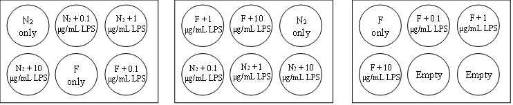
After adding the media, add the appropriate LPS dose to each well following the plate layout.
0.1 μg/mL LPS: 2 μL of 100 μg/mL LPS stock OR 20 μL of 10 μg/mL LPS stock
1 μg/mL LPS: 2 μL of 1 mg/mL LPS stock OR 20 μL of 100 μg/mL LPS stock
10 μg/mL LPS: 20 μL of 1 mg/mL LPS stock
Allow cells to incubate in the different treatment combinations for the required incubation time (3 hours).
After the required incubation time, remove the 6-well plates from the incubator and collect desired volume of media from each well.
Store media samples at -80°C for future use.
To perform TNF ELISA (using RayBiotech Rat TNF-alpha ELISA kit [Cat #ELR-TNFa-1, Norcross, GA]):
Bring all reagents/samples to room temperature.
Make the appropriate volume of 1X Diluent B by adding DI H2O to 5X Diluent B (Item E).
Standard Preparation:
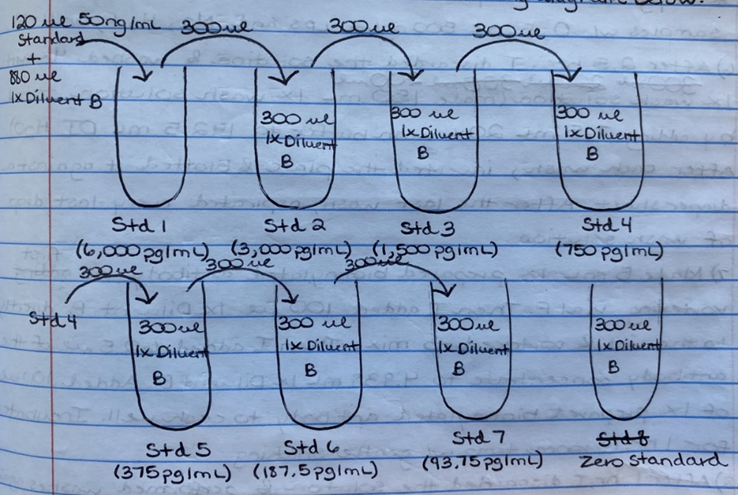
Gently vortex a vial of Standard Protein (Item C).
Add 400 μL 1X Diluent B directly to Item C and gently vortex to mix.
Add 120 μL diluted standard to 880 μL 1X Diluent B.
Perform a serial dilution to achieve the required standard concentrations (see example below).
Sample Preparation:
Dilute samples in 1X Diluent B.
For RT4-D6P2T media samples, use a 1:2 or 1:1 dilution.
1:2 dilution: 83.3 μL sample + 166.7 μL 1X Diluent B
1:1 dilution: 125 μL sample + 125 μL 1X Diluent B
Add 100 μL of each standard and sample into the appropriate wells of the 96-well plate following the plate layout (see example plate layout below).
| A | B | C | D | E | F | G | H |
|---|---|---|---|---|---|---|---|
| STD1 | STD1 | Sample 1 | Sample 1 | Sample 1 | Sample 9 | Sample 9 | Sample 9 |
| STD2 | STD2 | Sample 2 | Sample 2 | Sample 2 | Sample 10 | Sample 10 | Sample 10 |
| STD3 | STD3 | Sample 3 | Sample 3 | Sample 3 | Sample 11 | Sample 11 | Sample 11 |
| STD4 | STD4 | Sample 4 | Sample 4 | Sample 4 | Sample 12 | Sample 12 | Sample 12 |
| STD5 | STD5 | Sample 5 | Sample 5 | Sample 5 | |||
| STD6 | STD6 | Sample 6 | Sample 6 | Sample 6 | |||
| STD7 | STD7 | Sample 7 | Sample 7 | Sample 7 | |||
| ZERO STD | ZERO STD | Sample 8 | Sample 8 | Sample 8 |
Cover the plate and incubate for 2.5 hours on a rocker at room temperature.
While waiting, prepare the desired volume of 1X wash solution by adding DI H2O to 20X wash buffer.
Antibody Preparation:
Gently vortex the detection antibody (Item F).
Add 100 μL 1X Diluent B directly to the vial and gently pipette up and down to mix.
Dilute the detection antibody concentrate 80-fold using 1X Diluent B.
After 2.5 hours, discard the solution from each well.
Wash each well 4x with 1X wash solution. After each wash, invert the plate and blot it against a diaper sheet until the diaper sheet becomes dry. After the last wash, aspirate every last drop of wash solution.
Add 100 μL of the prepared detection antibody to each well, cover the plate, and incubate for 1 hour on a rocker at room temperature.
HRP-Streptavidin Preparation:
Gently vortex HRP-Streptavidin Concentrate (Item G) and pipette up and down to mix.
Dilute the HRP-Streptavidin Concentrate 200-fold using 1X Diluent B.
After 1 hour, discard the solution from each well and repeat washes following Step 10.
Add 100 μL of the prepared HRP-Streptavidin solution to each well, cover the plate, and incubate for 45 minutes on a rocker at room temperature.
After 45 minutes, discard the solution from each well and repeat washes following Step 10.
Add 100 μL TMB One-Step Substrate Reagent (Item H) to each well, cover the plate completely in aluminum foil, and incubate for 30 minutes on a rocker at room temperature in the dark.
After 30 minutes, add 50 μL Stop Solution (Item I) to each well. Immediately read the plate on the plate reader at 450 nm.
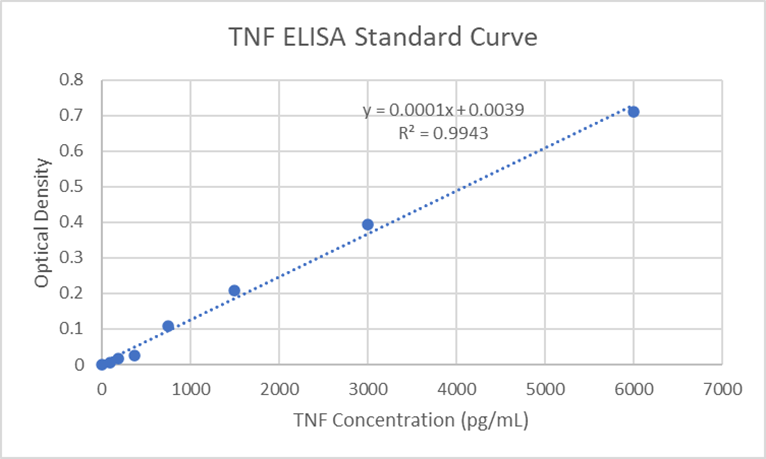
Plot the standard curve (see example below). Use the standard curve to approximate the concentration of TNF-alpha present in each sample. NOTE: Do not forget to correct for the dilution factor (for example, if you did a 1:1 dilution, multiply the TNF-alpha concentrations by 2).