Stem-cell-derived human microglia transplanted into mouse brain to study human disease
Nicola Fattorelli, Anna Martinez-Muriana, Leen Wolfs, Ivana Geric, Bart De Strooper, Renzo Mancuso
Published: 2021-01-10 DOI: 10.1038/s41596-020-00447-4


Extended
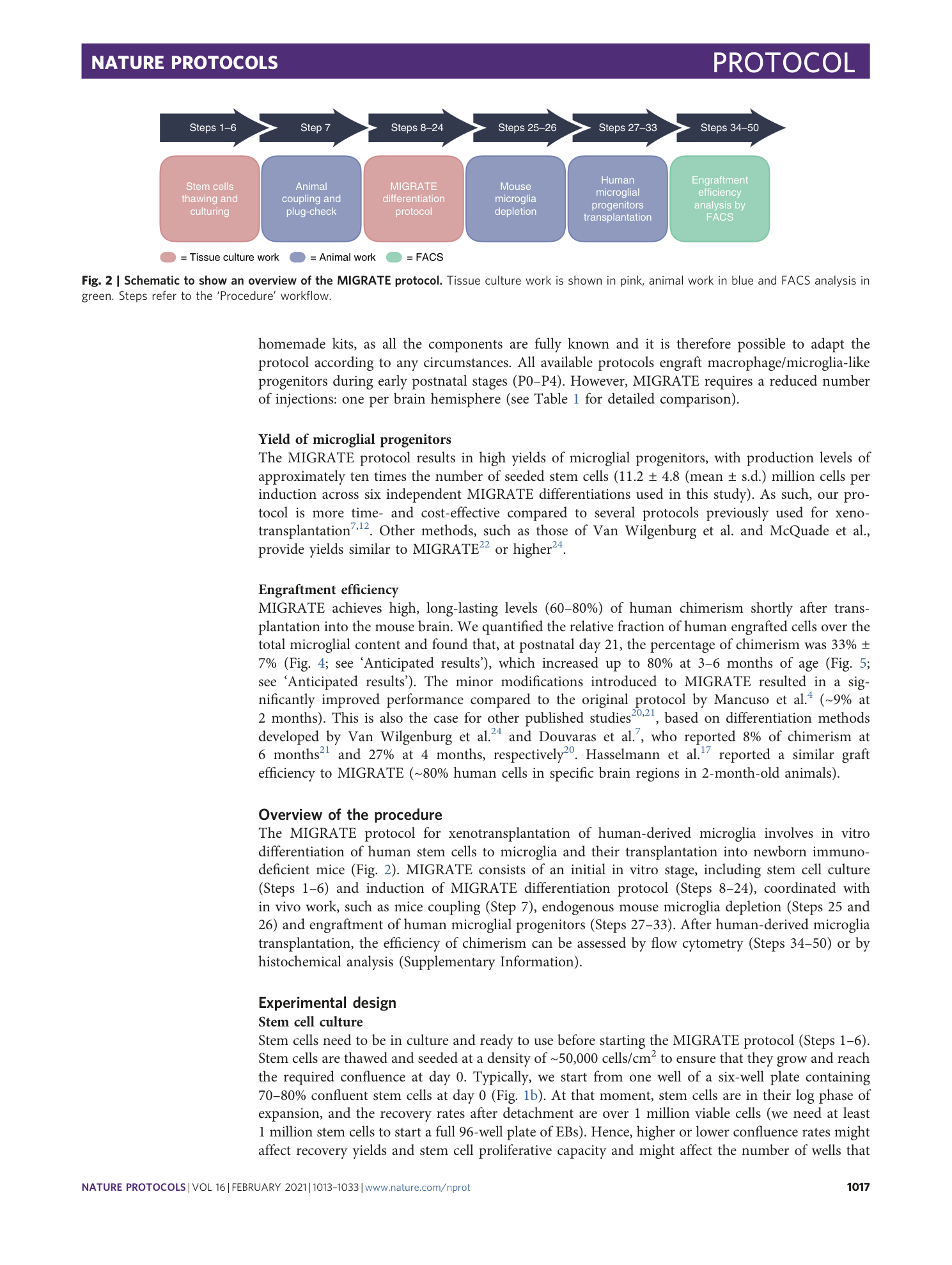
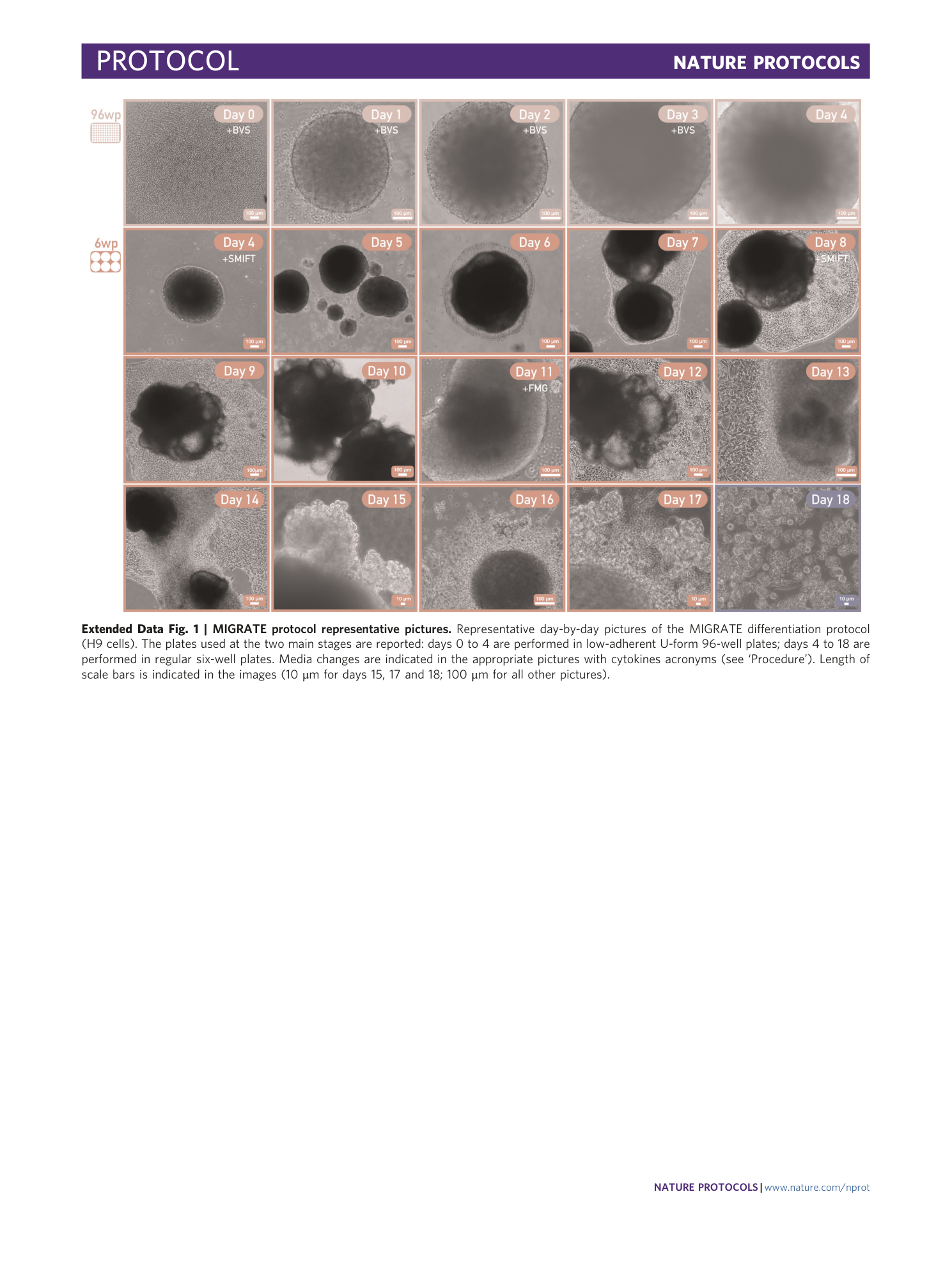
Extended Data Fig. 1 MIGRATE protocol representative pictures.
Representative day-by-day pictures of the MIGRATE differentiation protocol (H9 cells). The plates used at the two main stages are reported: days 0 to 4 are performed in low-adherent U-form 96-well plates; days 4 to 18 are performed in regular six-well plates. Media changes are indicated in the appropriate pictures with cytokines acronyms (see ‘Procedure’). Length of scale bars is indicated in the images (10 μm for days 15, 17 and 18; 100 μm for all other pictures).
Supplementary information
Supplementary Information
Supplementary Methods (histological analysis), Supplementary Tables 1 and 2 and Supplementary Fig. 1.
