Sonication of PFFs for use in vitro
Vijay Singh, Marta Castellana-Cruz, Nunilo Cremades, Laura Volpicelli-Daley
Disclaimer
DISCLAIMER – FOR INFORMATIONAL PURPOSES ONLY; USE AT YOUR OWN RISK
The protocol content here is for informational purposes only and does not constitute legal, medical, clinical, or safety advice, or otherwise; content added to protocols.io is not peer reviewed and may not have undergone a formal approval of any kind. Information presented in this protocol should not substitute for independent professional judgment, advice, diagnosis, or treatment. Any action you take or refrain from taking using or relying upon the information presented here is strictly at your own risk. You agree that neither the Company nor any of the authors, contributors, administrators, or anyone else associated with protocols.io, can be held responsible for your use of the information contained in or linked to this protocol or any of our Sites/Apps and Services.
Abstract
Animal models that accurately recapitulate the accumulation of alpha-synuclein (α-syn) inclusions, progressive neurodegeneration of the nigrostriatal system and motor deficits can be useful tools for Parkinson's disease (PD) research. The preformed fibril (PFF) synucleinopathy model in rodents generally displays these PD-relevant features, however, the magnitude and predictability of these events is far from established. We therefore have optimized the synthesis generation of α-syn fibrils to ensure reliable, robust results. These fibrils can be added to neurons in culture, differentiated iPSCs, or injected into mice or rats. The protocol includes steps for fibril synthesis as well as sonication for fibril fragmentaion which is a critical step for inducing formation of α-syn inclusions.
This updated protocol has been specified for use in vitro, and has been confirmed for use in both rat and mouse primary cultures.
Before start
Sonicating Fibril
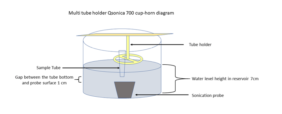
Proper sonication is a key step for the fibril model to work. For all in vivo work which involved injecting fibrils into mice or rats, we use the QSonica 700 sonicator with cup horn and tube rack for 1.5 mL polypropylene tubes with a chiller at 16°C. The cup horn sonication produces short fragments which maintain their morphology for 6-8 hours (at least) and can be stored in dry ice overnight, thawed and maintained at room temperature, and therefore remain active after overnight shipments.We found that over time, the heat generated by a probe tip sonicator causes the fibrils to form amorphous aggregates (Figure 1). This is a problem because stereotaxic surgeries can take several hours and the amorphous aggregates that form while the fibrils sit on the bench causes variability and reduces the concentration of seeding competent fragments. Another advantage of using the cup horn sonicator over probe tip is that 25 μL of fibrils can be sonicated, reducing the volume needed. This is also a closed tube system which increases safety. For neuron or cell culture work in which the fibrils are added to media and then the cells immediately after sonication, a probe tip sonicator is okay. Again, this should be performed in a BSL2 hood with all proper PPE (nanoparticle respirator, goggles, gloves etc.). The volume of fibrils to be sonicated cannot be less than 100 μL.
In all cases, we wear PPE when working with fibrils. We clean any spills with 1% SDS.

Attachments
Steps
PFF Sonication
Sonicator Info:
Equipment
| Value | Label |
|---|---|
| Q700 Sonicator | NAME |
| Sonicator | TYPE |
| QSonica | BRAND |
| Q700 | SKU |
| Q700 Sonicator with sound enclosure, cup horn, and recirculating chiller | SPECIFICATIONS |
Fill Qsonica water reservoir with about 900mL.
Turn on sonicator and recirculating chiller. Set the temperature at 10°C and allow to cool.
Thaw 30µL aliquots of PFFs at 0.200µg/µL at Room temperature
Transfer to
Total sonication time 0h 15m 0s
Pulse on 0h 0m 3s and off0h 0m 3s
Temperature 10°C
Amplitude at 45%
Sonication output should be between 90-100kJ
PFF Treatment
In culture hood, dilute PFFs to a volume of 300µL in 1x PBS for a concentration of 0.02µg/µL
Add 25µL (0.5µg) to each well of a 24-well plate. Equal volume 1x PBS was added for control conditions.
Age cultures for desired treatment length before fixation or collection.


