Sectioning Mouse Brain with Sliding Microtome
Naveen Ouellette, Holly Myers, daphne.toglia
Abstract
This is a protocol that details sectioning a mouse brain fixed in 4% PFA and 30% sucrose on a sliding microtome. This protocol details the microtome set up, the mounting of the brain on the microtome stage in OCT or 30% sucrose, slicing the brain with the microtome blade, microtome take down and clean up, and tissue section storage.
Before start
The mouse brain must be fixed in 30% sucrose for 1-2 days prior to sectioning on microtome, or until the brain sinks to the bottom of the container of 30% sucrose.
Steps
Setup
Work station setup:
Set up a well plate next to the microtome. This will be used to store the brain sections that are sliced during this protocol. Wells should be filled with 1xPBS & Sodium Azide 0.01% for tissue preservation. Label the well plate lid with relevant information: brain ID number, section thickness, date, etc.
Set up a dish filled with 1xPBS next to the microtome to collect any miscellaneous sliced tissue that will be discarded and not saved for storage.
Microtome setup:
Microtome diagrams with labeled parts:
<img src="https://static.yanyin.tech/literature_test/protocol_io_true/protocols.io.5jyl8p97rg2w/p6xcb578714.jpg" alt="Left side of microtomeA - knobs adjust angle of stageB - stageC - knifeD - knife clampsE - knife guard - slides left to cover knifeF - lever locks stage in placeG - coarse feed wheel - raises stage up and downH - manual feed lever - raises stage up by amount of microns set with dial "I"I - section thickness adjustment dial (up to 60 microns)" loading="lazy" title="Left side of microtomeA - knobs adjust angle of stageB - stageC - knifeD - knife clampsE - knife guard - slides left to cover knifeF - lever locks stage in placeG - coarse feed wheel - raises stage up and downH - manual feed lever - raises stage up by amount of microns set with dial "I"I - section thickness adjustment dial (up to 60 microns)"/>
L - number indicate angle of knifeM - knife angle adjustment knob")
Ensure lock handle for knife is in the closed position before making any adjustments to, or mounting tissue onto, the microtome.
<img src="https://static.yanyin.tech/literature_test/protocol_io_true/protocols.io.5jyl8p97rg2w/p6vub57871.jpg" alt="Knife guard is locked when the handle is up. See "K" in diagram in step 2.1." loading="lazy" title="Knife guard is locked when the handle is up. See "K" in diagram in step 2.1."/>
<img src="https://static.yanyin.tech/literature_test/protocol_io_true/protocols.io.5jyl8p97rg2w/p6vyb57872.jpg" alt="Knife guard is unlocked when the handle is down. See "K" in diagram in step 2.1." loading="lazy" title="Knife guard is unlocked when the handle is down. See "K" in diagram in step 2.1."/>
Attach the stage to the microtome and ensure that it is level.
<img src="https://static.yanyin.tech/literature_test/protocol_io_true/protocols.io.5jyl8p97rg2w/p6v4b57873.jpg" alt="Pictured is a level tool sitting on top of the metallic microtome stage. The angle of the stage can be adjusted from a side knob (see "A" in step 2.1) until the level tool confirms that the stage is level when the bubble is in the center/bulls-eye of the circles." loading="lazy" title="Pictured is a level tool sitting on top of the metallic microtome stage. The angle of the stage can be adjusted from a side knob (see "A" in step 2.1) until the level tool confirms that the stage is level when the bubble is in the center/bulls-eye of the circles."/>
Take microtome knife out of its case and use 70% ethyl alcohol and a Kimwipe to remove the protective oil coating on the knife.
Insert knife into the holder and tighten both black-knobbed clamps to hold knife in place. Black knife guard located below knobs can be used to cover blade when microtome blade is inserted but not in use.
<img src="https://static.yanyin.tech/literature_test/protocol_io_true/protocols.io.5jyl8p97rg2w/p6v8b57875.jpg" alt="Metal microtome blade installed into microtome ("C", step 2.1). Two knife clamps with black knobs located to right of blade ("D", step 2.1), and black plastic knife guard located below clamps slides left to cover knife ("E", step 2.1)." loading="lazy" title="Metal microtome blade installed into microtome ("C", step 2.1). Two knife clamps with black knobs located to right of blade ("D", step 2.1), and black plastic knife guard located below clamps slides left to cover knife ("E", step 2.1)."/>
Set the knife angle to desired setting with knife angle adjustment knob. For example, 0 degrees.
<img src="https://static.yanyin.tech/literature_test/protocol_io_true/protocols.io.5jyl8p97rg2w/p6wcb57876.jpg" alt="Knife angle adjustment knob located on top of microtome (see "M", step 2.1 diagram) and numbers indicating knife angle located on right side of microtome (see "L", step 2.1 diagram). " loading="lazy" title="Knife angle adjustment knob located on top of microtome (see "M", step 2.1 diagram) and numbers indicating knife angle located on right side of microtome (see "L", step 2.1 diagram). "/>
Adjust the section thickness in microns by turning the section thickness adjustment dial.
<img src="https://static.yanyin.tech/literature_test/protocol_io_true/protocols.io.5jyl8p97rg2w/p6wgb57877.jpg" alt="Dial located on the front of the microtome measures section thickness in microns. ("I" in step 2.1 diagram) Dial is set to 50 microns in this image." loading="lazy" title="Dial located on the front of the microtome measures section thickness in microns. ("I" in step 2.1 diagram) Dial is set to 50 microns in this image."/>
Create a base of 1xPBS, OCT, or 30% sucrose on the microtome stage:
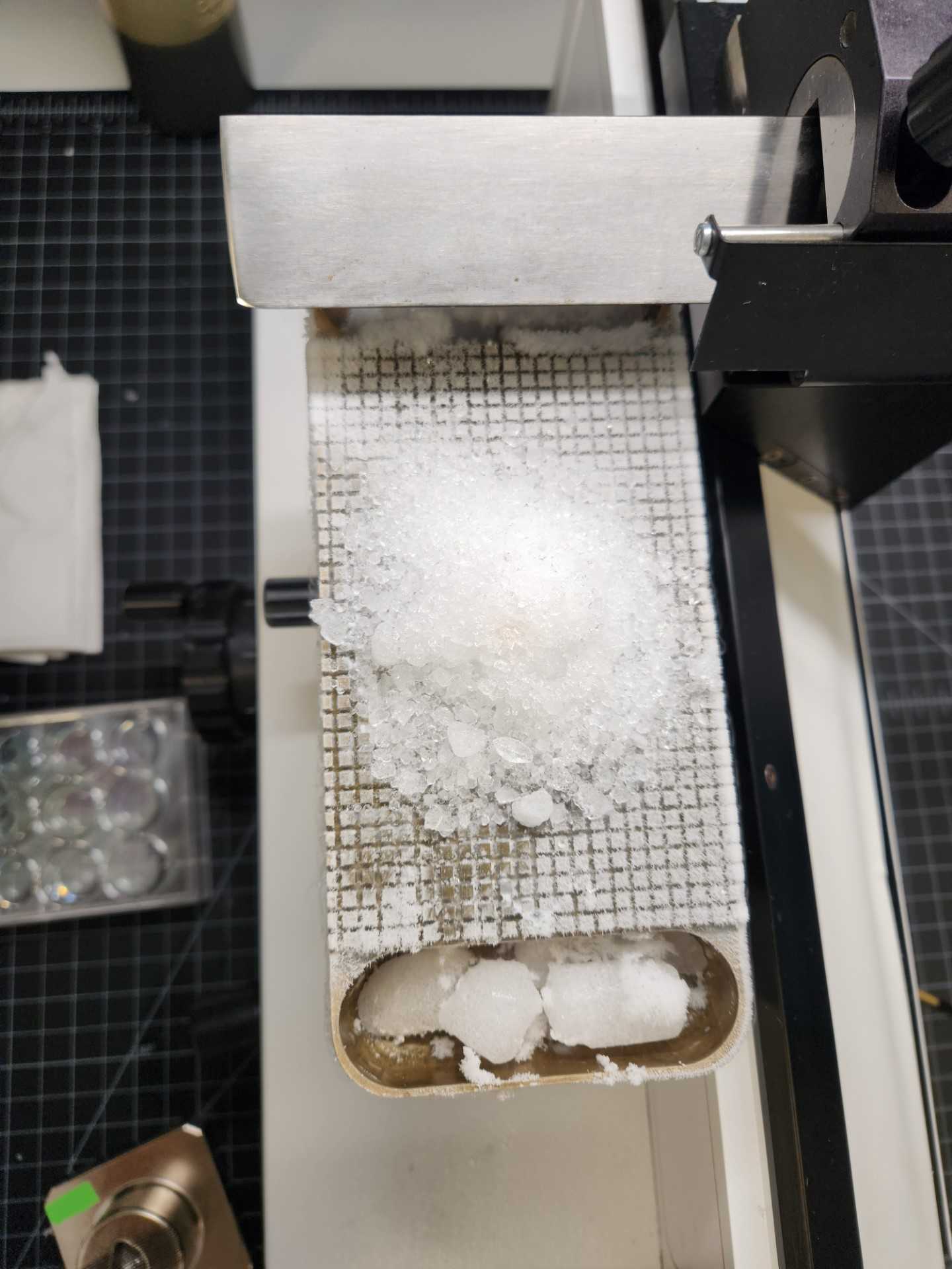
Fill the trough surrounding the stage with dry ice chunks.

Wait until stage has cooled enough so that the 1xPBS, OCT, or sucrose will freeze soon after it is applied. The metallic stage should look slightly white and frosty, and might take 0h 5m 0s to reach this point.
Apply a 4cm square layer of 1xPBS on the stage to section one brain.


Wait until the 1xPBS is completely frozen before proceeding.
Determine if you will mount the specimen in OCT or sucrose. If mounting in OCT, proceed to step 4. If mounting in sucrose, proceed to step 5.
| A | B |
|---|---|
| OCT | -Quicker process to mount the specimen. |
| 30% sucrose | -Easier to cut through with a knife. -Good for larger specimens like macaque brain. -Easier to replace remaining specimen back in 30% sucrose for storage for later use. |
Mounting specimen using OCT
Mount the specimen on microtome stage using OCT:
With a razor blade, trim tissue as needed so it will sit flat and level on the stage in the correct orientation (coronal, sagittal) when sectioning.
Blot specimen briefly on a Kimwipe to remove excess liquid.
Add a large drop of OCT onto the frozen, flat OCT base.
Quickly set the specimen into the large drop of OCT before it freezes.
Gently hold the tissue in place until the OCT has solidified enough to hold the tissue securely.

Crush dry ice with a rubber mallet and apply ice dust to the top of the mounted brain.
Wait until tissue freezes passively, about 0h 1m 0s , and gently brush away crushed ice to reveal brain specimen. The tissue should be hard and white after freezing.
Proceed to step 6 for sectioning.
Mounting specimen using 30% sucrose
Mount the specimen on microtome stage using 30% sucrose:
With a razor blade, trim tissue as needed so it will sit flat and level on the stage in the correct orientation (coronal, sagittal) when sectioning.
Blot specimen briefly on a Kimwipe to remove excess liquid.
Add a large drop of 30% sucrose onto the frozen, flat 30% sucrose base.
Quickly set the specimen into the large drop of sucrose before it freezes.
Surround base with more sucrose until you have a sturdy base that will hold the specimen securely. Add slowly drop by drop and avoid making hollow cavities.
Keep adding sucrose until specimen is securely held or if preferred, completely
covered with sucrose.

Sectioning
Section the tissue:
If desired by requestor, use a razor blade to cut a shallow notch on either the left or right side of the cortex of the brain that runs posterior to anterior. Make a note of which side is notched and refer to it for left/right orientation when mounting sections on slides.
Add more dry ice as needed to the troughs in the stage to prevent the OCT or sucrose mounting solution from softening or melting, as the dry ice added initially will melt over time.
Repeat steps 6.4 - 6.10 until desired sections are collected. Tissue may be double or triple stacked in each well in the well plate, but ideally sections in the same well should be separated by at least a few hundred microns (ex: every 6th section or more) so it is easier to see anatomical differences and determine section order when mounting.
Wrap well plate in parafilm and secure with tape to prevent spillage, then wrap in aluminum foil to protect from light, and store at 4°C until ready to stain the tissue and mount the sections onto slides. Tissue may be stored for several months.
Move the platform and specimen vertically towards the knife by turning the coarse feed wheel located on the left side of the microtome.
To position blade as close as possible to the specimen, pull the manual feed lever forward to raise the platform by microns.
Pull the knife sledge movement grip forward to bring the knife through the tissue.
The tissue section will remain on the top of the knife. Wrinkles and deformation of the tissue is normal.
Pick up the section with a fine-tipped paintbrush. Move each section to the well plate filled with 1xPBS & Sodium Azide 0.01% .
If needed, wipe the blade with a Kimwipe to clear any excess moisture or debris.
Push the knife sledge movement grip back to bring the blade back to the starting point.
Pull the manual feed lever forward to raise the specimen for the next section.
Clean up
Take down microtome and clean up equipment:
Spray the knife and microtome with 70% ethyl alcohol and carefully wipe dry with Kimwipes.
Coat the knife with mineral oil and carefully store in its case.
Detach the specimen stage and run warm tap water over it to release 1xPBS, OCT, or 30% sucrose and any residual tissue.
Based on requestor preference, either retain tissue or dispose of tissue in biohazard waste bin.
Spray the stage with 70% ethyl alcohol. Soak for 0h 3m 0s before wiping with a Kimwipe.
Dry the stage thoroughly to prevent frost buildup.
