Quick DNA Extraction for Fungal Barcoding (X-Amp)
Stephen Douglas Russell
Abstract
Below you will find a simple and fast two-step DNA extraction protocol that works well with many different groups of fungi. This protocol can be utilized in preparation for DNA barcoding of fungi utilizing Oxford Nanopore MinION.
Time required:
Varies depending on the use case. My samples generally have the metadata and images stored on iNaturalist. For each sample, I go to the iNaturalist observation, validate that the specimen matches the metadata and images, include a unique lab code in the "Voucher Number(s)" observational field, record the lab code on the sample packet with a marker, and record the lab code on the associated spreadsheet in the correct cell number.
If all of these actions need to be performed, it can take 3-5 hours per 96 samples to load.
If the iNaturalist observations have already been validated, the lab numbers have been assigned, and all that is required is to put a small amount of tissue in each cell for each sample, then the time outlay can be less than 2 hours per 96 samples.
Limitations on Protocol:
There are a number of species groups that will have lower success rates with this protocol. They will either need a more complex DNA extraction protocol that involves grinding, or different primer combinations.
Low success rates (0-20% of specimens): Cantharellus, Helvella, Strobilomyces, Ophiocordyceps
Moderate success rates (20-60% of specimens): Recalcitrant crusts & polypores, Cuphophyllus, Neohygrocybe, Tylopilus
Steps
Initial Preparation
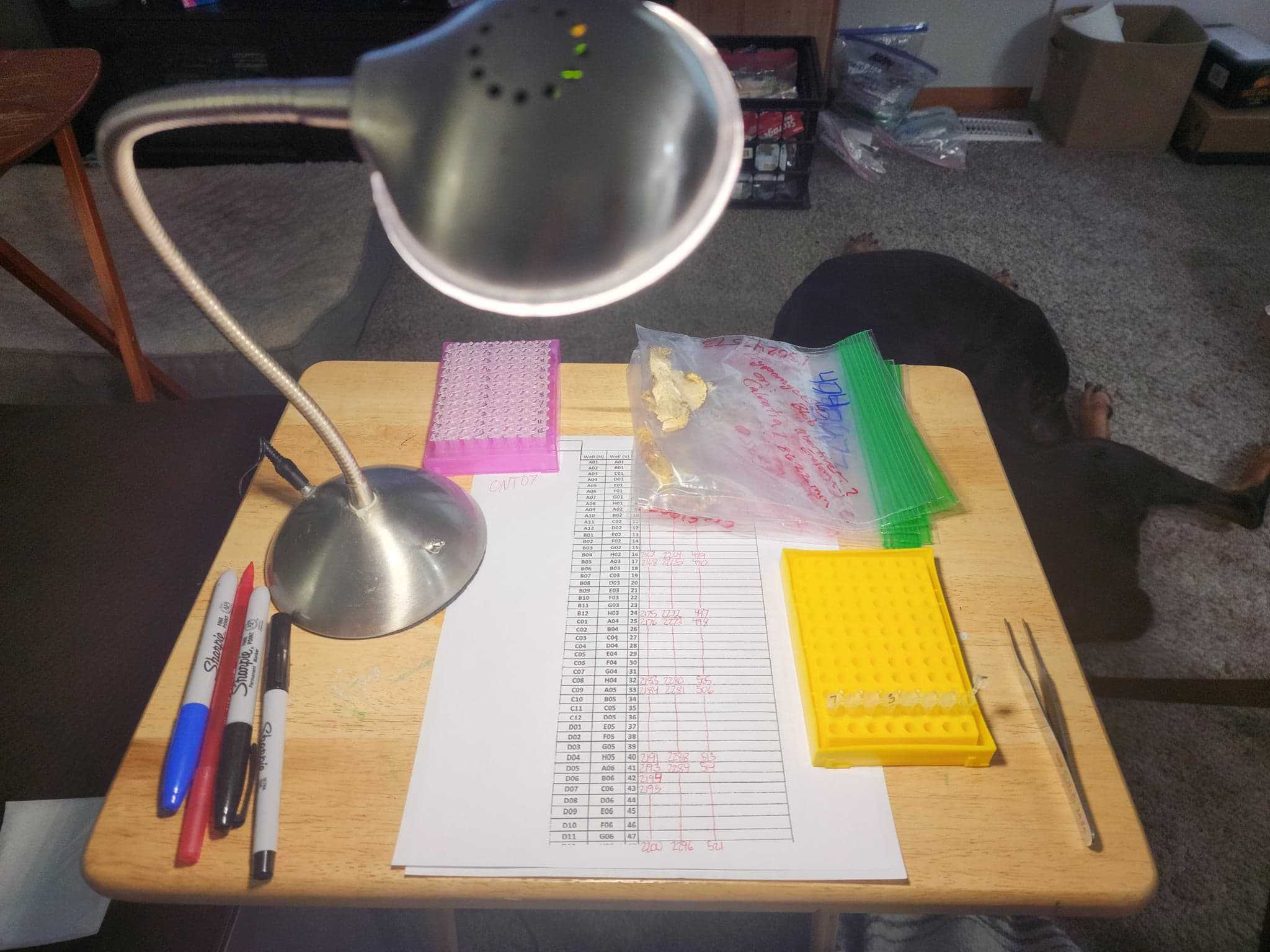
Begin by laying out (96) 0.2mL cells on a 96-well PCR plate rack. This can be done in a plate or strip format.
Even though it costs more, I prefer to use 8-strips with individual caps as they are easier to work with. Only a single sample cell needs to be open at a time, individual rows can be removed for closer examination, spin-down at the end of extraction is easier (vs. plates), etc.
Label the cells with the Run #, the plate #, and the row number.
<img src="https://static.yanyin.tech/literature_test/protocol_io_true/protocols.io.4r3l2oy2pv1y/jhkfbtupp1.jpg" alt="Above is plate number 2 on run number "ONT07." Plates are filled from right to left. I usually work with two plate racks, one holding the strip I am currently working on and the other holding the remainder of the filled and empty tubes. " loading="lazy" title="Above is plate number 2 on run number "ONT07." Plates are filled from right to left. I usually work with two plate racks, one holding the strip I am currently working on and the other holding the remainder of the filled and empty tubes. "/>
I prefer a physical printout for keeping a record of the sample-cell associations. I use the sheet linked below. You could also keep track digitally on a laptop or computer if that is your preference.
Before starting, I will typically put the specimens in numerical order, which includes adding my own lab/herbarium code to the specimens. This makes the final digital spreadsheet preparation much easier. Having the samples in order allows the final spreadsheet to be populated by drag-and-drop in Excel and it allows all of the iNat numbers to be copy and pasted (always with some minor modification) directly into the final sample spreadsheet.
As an example, I will lay out all of my samples, mark the bags with a lab code (ex - 1 to 96) and enter the same lab code into the iNat Voucher number(s) observational field (Ex - OSF2022-096). I then pull tissue from the samples in that order, filling the cells from right to left across the strip. (Loading right to left allows the flow to go from A -> H of a typical 96-well plate style format.)
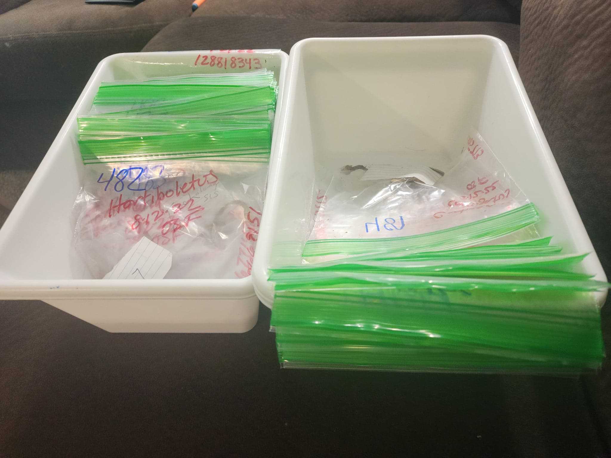
This takes some time initially, but it will save time with later with spreadsheet preparation. It typically takes me 15-30 minutes to fully populate the final spreadsheet with this prep-work. It would otherwise take several hours.
The ultimate methodology you utilize will depend on the types of samples you are processing, the format of the voucher numbers, and the location of the metadata/images.
X-Amp Extractions
Add a small amount of fungal tissue to each cell. The amount of tissue should be small enough to easily drop to the bottom of the tube. Less is better than more. If you can physically see the sample in the tube it is probably enough for a successful extraction. Remember, you will be filling the tubes with tissue from right to left.
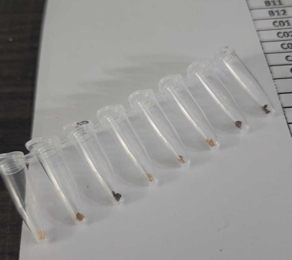
For dried specimens, I will typically break off a small section of the mushroom with my fingers and place it on a Kimwipe, blank white paper, or on the field data slip in the bag. From there it can be further broken up until you spot a piece of the desired size to put in the tube.
For fresh specimens, I will try to pull off the desired amount of tissue for each cell directly from the specimen. Note - I no longer use fresh specimens for this protocol. They are more difficult to work with.
Both dried and fresh specimens use the same visual amount of tissue. Dried tissue is MUCH easier to work with than fresh tissue in these small tubes, as the fresh tissue tends to stick to unintended places.
Between specimens, just wipe them off quickly with a Kimwipe. No need to flame sterilize or to utilize alcohol.
Place 15µL to 25µL of 20µL. Using less solution means the overall cost basis per sample will be lower. This process is easiest with a multichannel pipette. One option is to keep a PCR Rack dedicated to an 8-strip of X-Amp for pipetting.
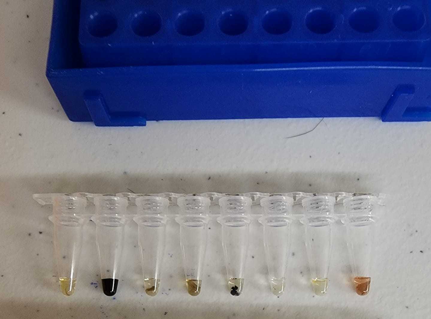
Once all of the strips on the plate have X-amp, tap down each strip with your finger to ensure there are no air bubbles in the bottom of the tube underneath the liquid and that the tissue is completely submerged in the fluid. Spin down each strip for 10 seconds in a mini centrifuge. As you pull them out, once again, doublecheck to ensure the tissue in each cell is in the fluid. It is ok if a sample is too large for the amount of fluid and only partially submerged.
Heat the tubes with the tissue for 1h 0m 0s at 80°C in a thermocycler.
Add 100µL of 100µL.
I utilize two PCR racks with the 8-strips every other row for this process. It makes opening and closing flip-top tubes easier. It is less of a consideration for strip caps.
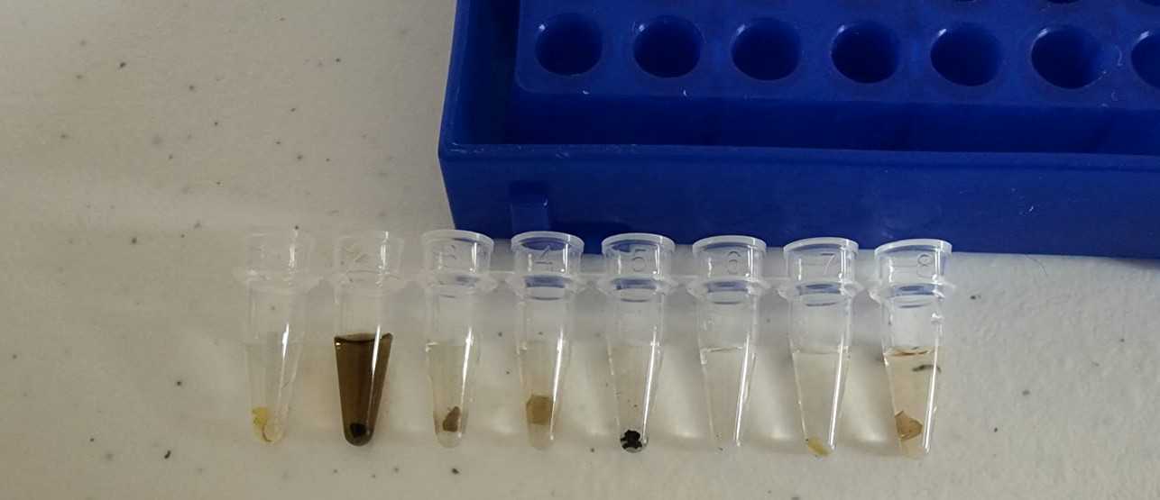
Optional: Microwave the samples for 2 minutes. I typically do this as a part of the protocol. In case you are not familiar, there are a number of microwave-only DNA extraction protocols. I look at this final step as a "thermal shock" heating, which disrupts the cell wall more than a standard heat bath.
This template is now ready for use in PCR reactions.

