Protocol for detection of Salmonella Typhi and Salmonella Paratyphi A in Surface Swabs
Renuka Kapoor, Christine Moe
surface
tile
wood
metal
swab
Salmonella Typhi
Salmonella Paratyphi A
enrichment
detection
environment
Abstract
The protocol describes method for qualitative detection (presence/ absence) of Salmonella Typhi and Salmonella Paratyphi A from surface swabs by enrichment culture followed by real-time PCR.
Steps
1. Processing of surface swab samples
The following steps describe processing of surface swabs up to enrichment stage.
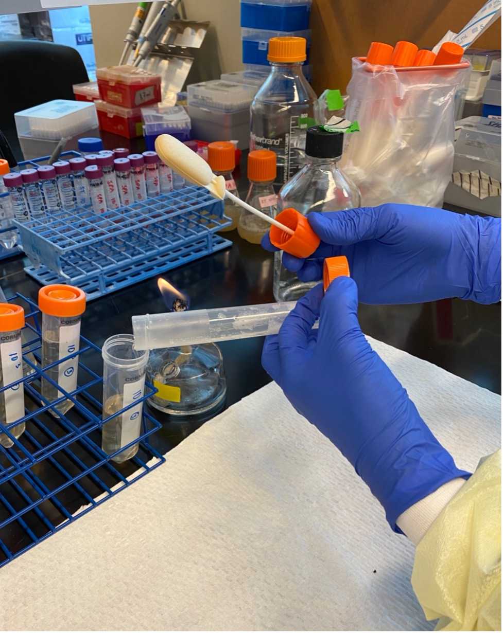
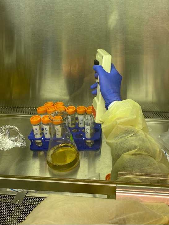
Put on gloves and spray hands with 70% ethanol and rub hands together to sanitize all surfaces of the gloves.
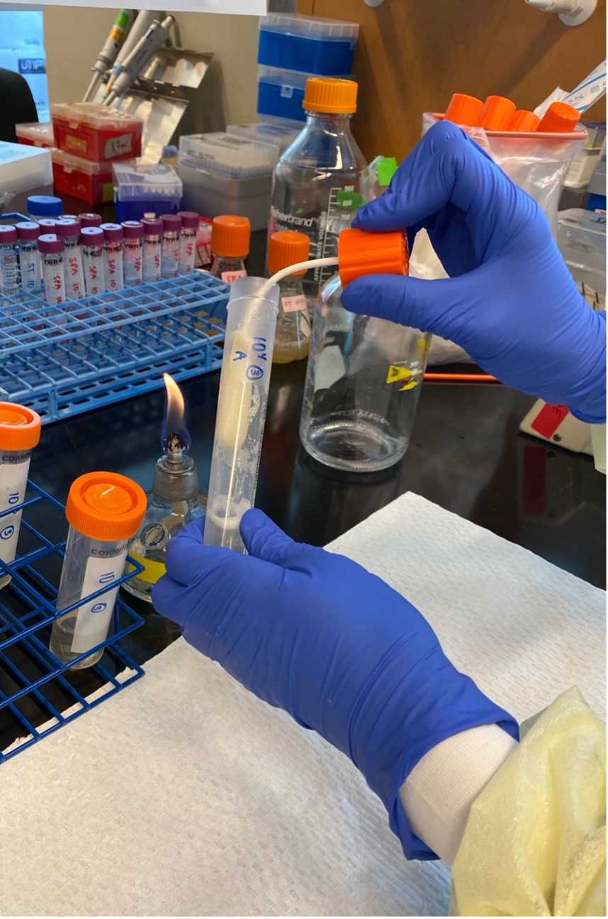
Reopen swab container and pour the swab elute into the empty labeled 50 mL conical tube.
Wash the swab again with 10 ml of PBST by repeating steps 1.5 through 1.9, then proceed to step 1.12.
Press the swab against the side of the tube to squeeze out as much remaining wash solution as possible, and transfer into the 50 mL conical tube.
Prepare your work surface by cleaning it with 70% ethanol.
Label a sterile 50 mL conical tube each with the Sample ID, the date, and your initials.
Carefully unscrew the cap of the tube with the swab.
Use a sterile 10 mL pipet to carefully add 10mL of PBST to the swab containers.
Place the swab back into the tube, screwing the lid down well.
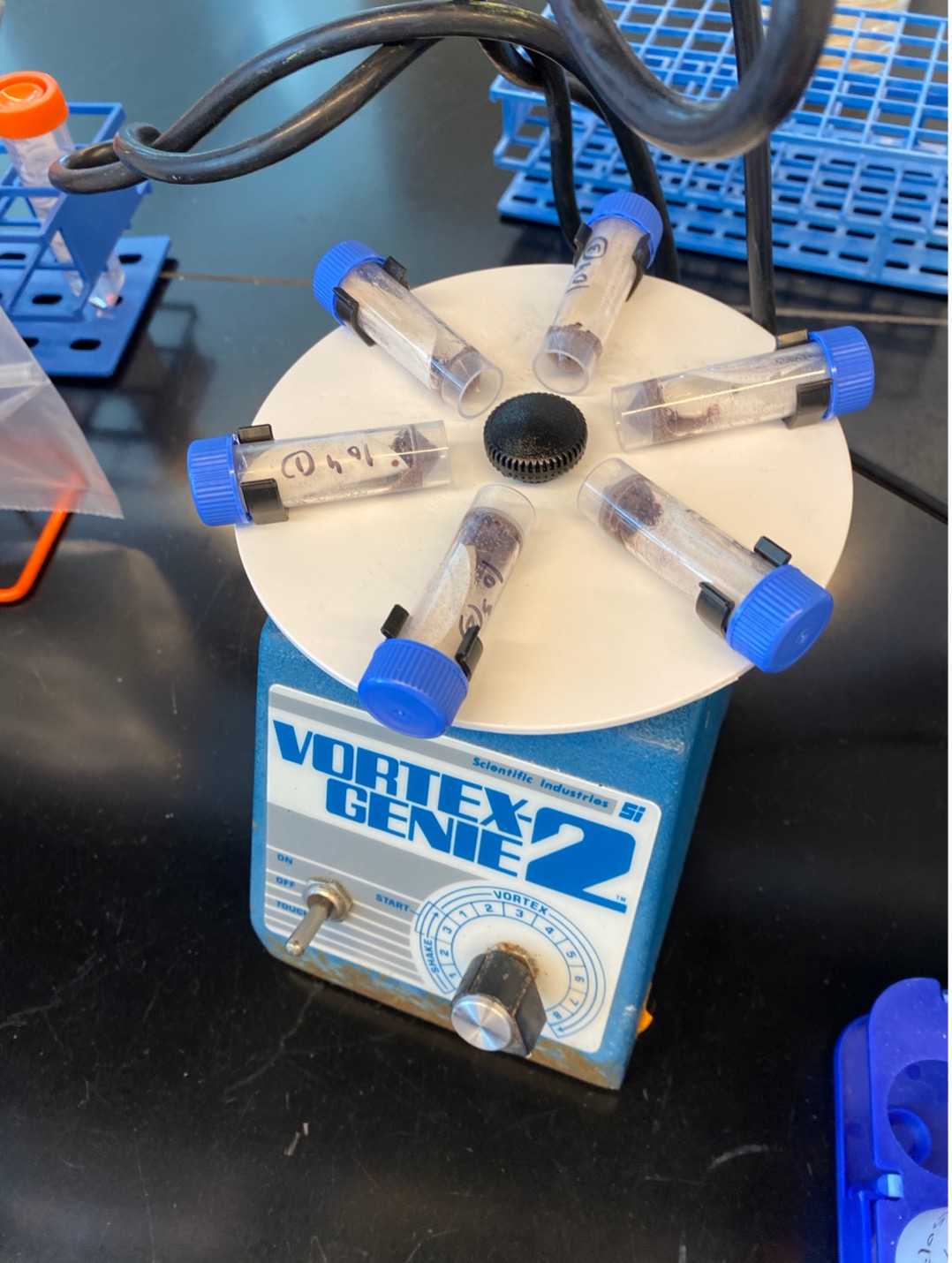
Vortex the tubes containing the swabs for 0h 0m 30s.
Incubate for 0h 5m 0s at Room temperature.
Vortex for another 0h 0m 30s.
2. Enrichment culture
The following steps are for optional enrichment of the sample. If enrichment is not performed, the processed sample can be used directly for membrane filtration and DNA extraction (Section 3).
In a biosafety hood, transfer 180mL of Universal Pre-enrichment (UP) Broth (USEPA Standard Analytical Protocol for Salmonella Typhi in Drinking Water) to a sterile 500 mL flask.
Add 10mL of swab elute to the flask containing UP Broth.
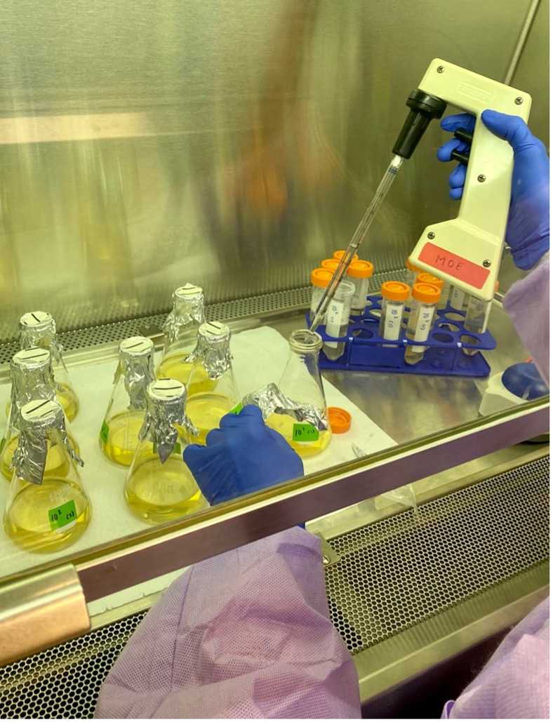
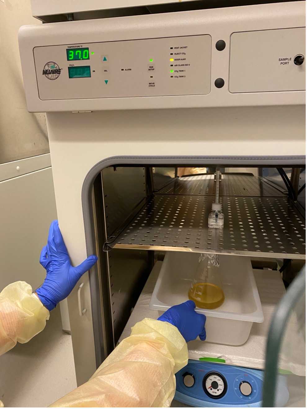
Incubate the flask in shaking incubator 37°C overnight.
3. DNA Extraction
The following steps are for membrane filtration and DNA extraction. They can be performed following step 1.12 or step 2.3.
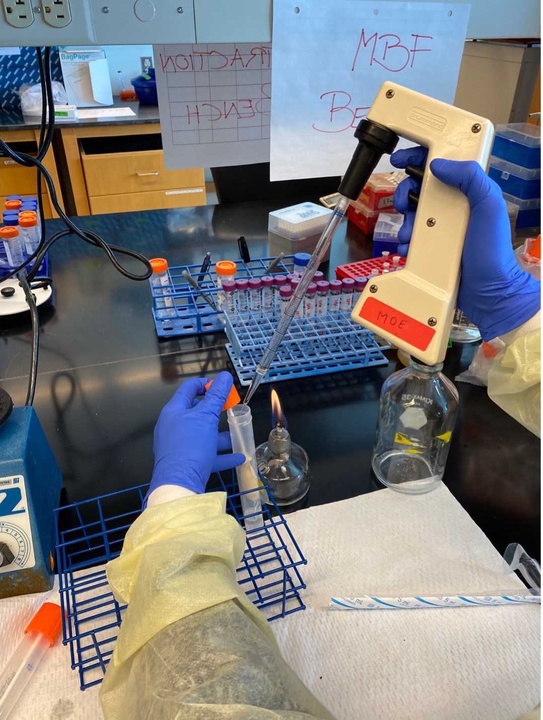
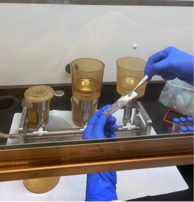
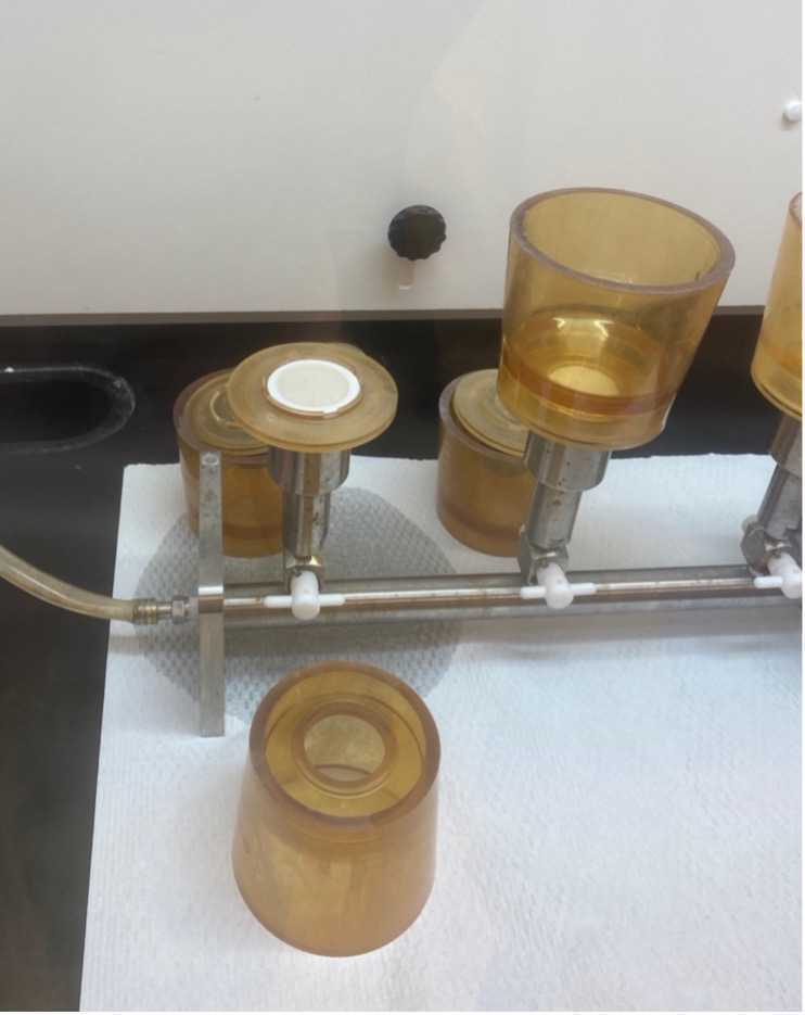
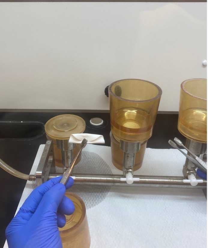
Clean your workspace and set up your filter units.
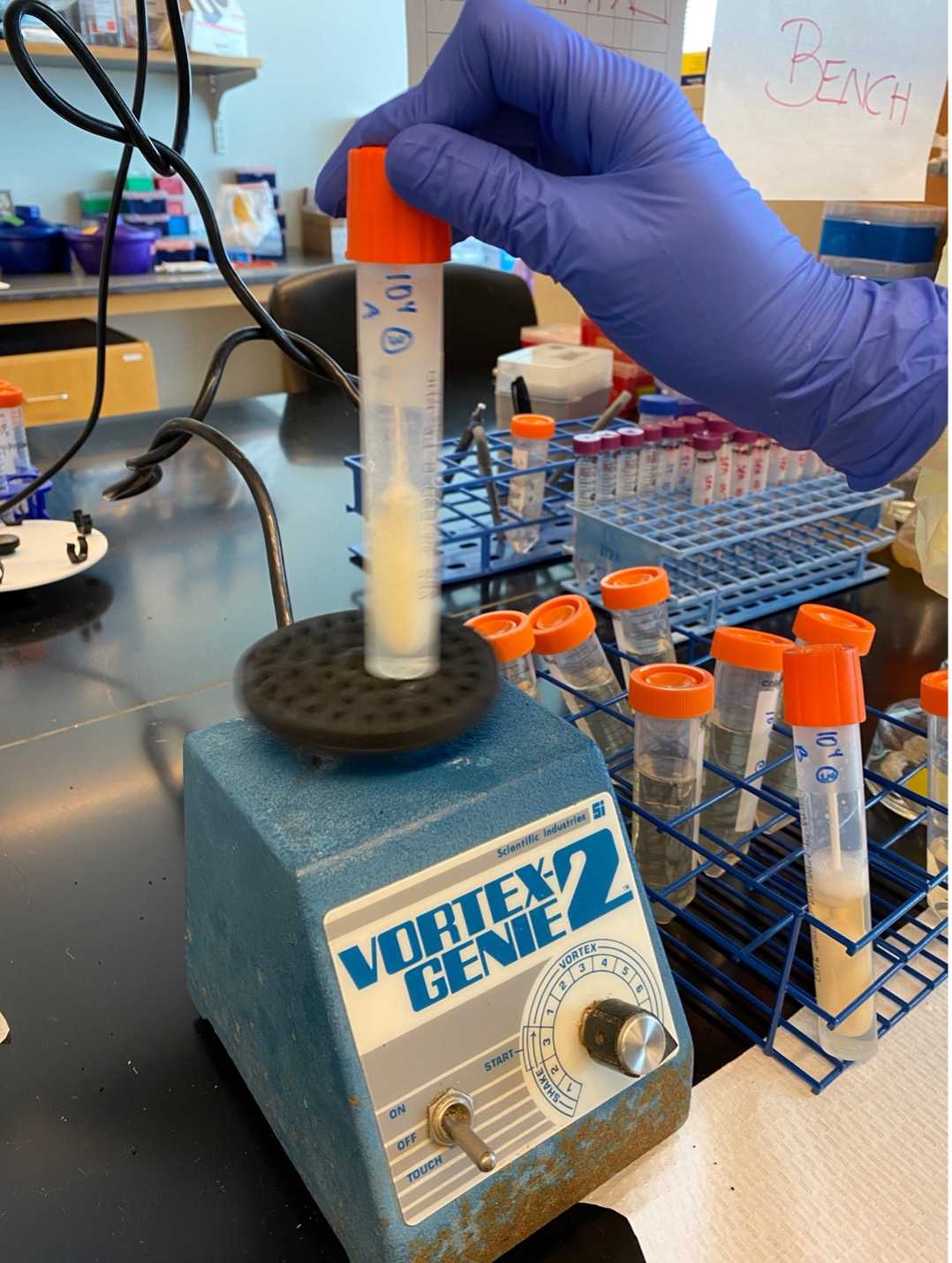
Transfer the folded filter to a bead tube (from Qiagen DNeasy PowerWater kit).

Add 1mL of Buffer PW1 (Qiagen DNeasy PowerWater kit) to the bead tube and vortex for 0h 5m 0s.
Proceed with the DNA Extraction according to manufacturer’s protocol (Qiagen DNeasy PowerWater kit).
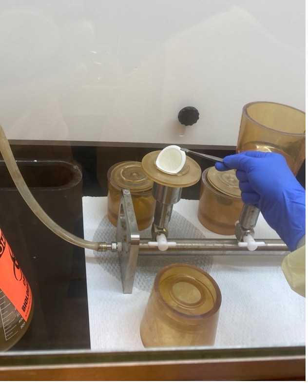
Using sterile forceps, place a clean membrane filter on the base of the filter unit.
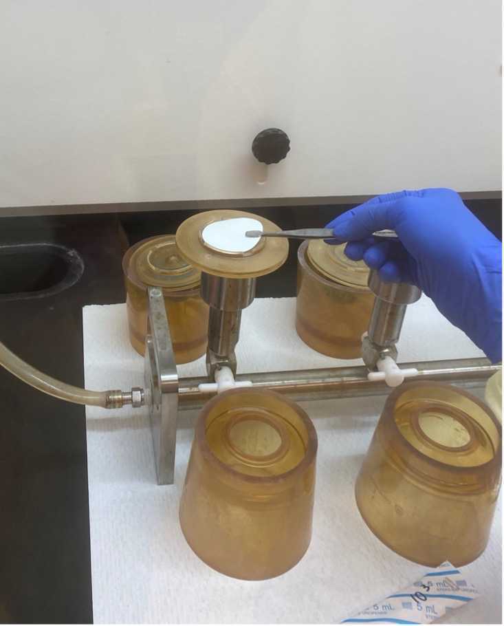
Place the cup on top of the filter.
Add 20mL of enriched sample to the cup and turn on the vacuum.
Allow the sample to filter until liquid is no longer visible on the filter.
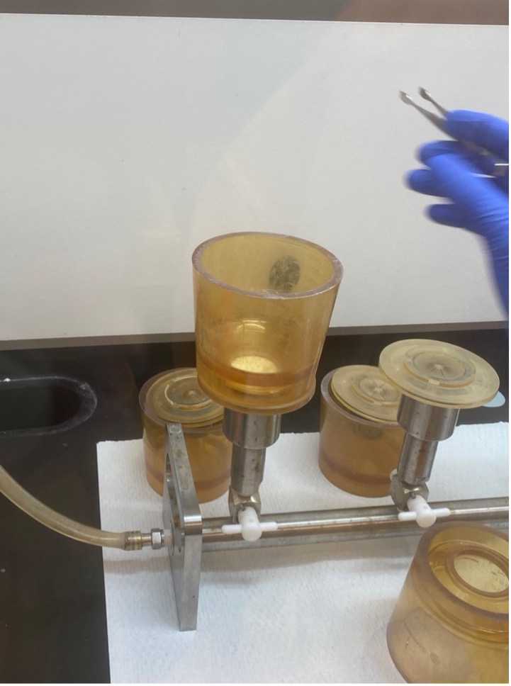
Turn off the vacuum and remove the cup from the base.
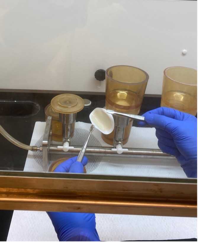
Using a sterile forcep, remove the filter from the base.
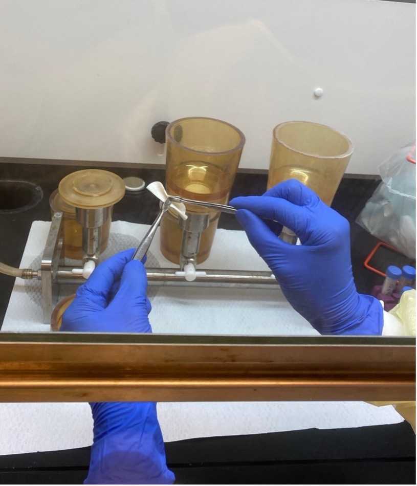
Using two sterile forceps, fold the filter in half inward, so the cells are now contained inside the folded filter.

Fold the filter in half again.
4. Real-time PCR
Test DNA extracts for S . Typhi and S . Paratyphi A using Taqman-based quantitative real-time PCR (qPCR) platform.
Detection of S . Typhi
S . Typhi is detected using duplex PCR protocol developed by researchers at the University of Washington (Scott Meschke and team) usIng primers and probes targeting the tviB and staG genes (Nair et al., 2019).
tviB _F 5’TGTGGTAAAGGAACTCGGTAAA-3’;
tvi B_R 5’-GACTTCCGATACCGGGATAATG-3’;
tvi B_P HEX - TGGATGCCGAAGAGGTAAGACGAGA-BHQ1;
sta G ___ F 5’- CGCGAAGTCAGAGTCGACATAG-3’;
sta G ___ R 5’-AAGACCTCAACGCCGATCAC-3’;
sta G ___ P FAM-5’-CATTTGTTCTGGAGCAGGCTGACGG-3’-BHQ1
The reaction mixture contain 0.65 µl of tviB_F (20µM), 0.75 µl each of tviB_R (20µM), staG_F(20µM),and staG_R (20µm), 0.5 µl each of the probe tviB_P (10µM) and staG_P (10 µM), 12.5 µl of SsoAdvanced Universal Probes Supermix (Bio-rad), and 5 µl of DNA in a final volume of 25 µl. The PCR reaction conditions include initial denaturation at 95oC for 5 min, followed by 45 cycles of 95oC 30 sec, 64oC 30 sec, 72oC 10 sec, and final extension at 72oC for 5 min.
Detection of S . Paratyphi A
S . Paratyphi A Is detected using primers and probe targeting SPA2308 (Nga et al., 2010)
SPA2308_F 5’- ACGATGATGACTGATTTATCGAAC-3’;
SPA2308_R5’-TGAAAAGATATCTCTCAGAGCTGG-3’;
SPA2308_PCY5 - CCCATACAATTTCATTCTTATTGAGAATGCGC-BHQ2
The reaction mixture containing 1 µl of each primer (10µM), 0.4 µl of probe (10µM), 200µM of dNTPs, 5mM of MgCl2, 5U of HotStar Taq DNA polymerase (Qiagen), and 5 µl of DNA in a final reaction volume of 25 µl. The PCR reaction conditions include initial denaturation at 95oC for 5 min, followed by 45 cycles of 95oC 30 sec, 60oC 30 sec, 72oC 30 sec, and final extension at 72oC for 10 min.
