Phenocycler-Fusion Staining Protocol For Bone Marrow Tissue
Kyung J Ahn, Shovik Bandyopadhyay, Anusha Thadi, Kai Tan
Abstract
This protocol describes the method for antibody staining of FFPE bone marrow tissue on slides using CODEX barcoded antibodies. The Akoya Phenocycler-Fusion user manual was modified to include photobleaching steps. Included are the stepwise protocols for tissue procurement, tissue decalcification, tissue sectioning, pre-staining, deparaffinization, antigen retrieval, photobleaching, antibody staining, post-fixation, and tissue storage.
Steps
Tissue Procurement
Femoral head bone marrow tissue obtained from total hip arthroplasty surgery was cut coronally in 4-6 mm thick slices by the Anatomical Pathology Lab at Penn Presbyterian Medical Center.
A dental biopsy drill was used to harvest 4 mm diameter cylindrical bone marrow tissue samples.
Tissue Fixation
Bone marrow tissue was fixed in 4% paraformaldehyde at 4°C for 24 hours.
Tissue Decalcification
Bone marrow tissue was washed 3 times for 30 minutes in PBS at 4°C.
Bone marrow tissue was decalcified in 0.5M EDTA pH8 for 8 days at 4°C. The EDTA was changed every 2 days.
After decalcification, samples were washed 5 times for 1 hour in 1 X PBS at 4°C.
Bone marrow tissue was fixed overnight in 4% paraformaldehyde at 4°C.
FFPE Blocks
Bone marrow tissue was processed and embedded in paraffin by the Children’s Hospital of Philadelphia pathology core.
FFPE Tissue Sectioning
Prepare a water bath at 40°C.
Section the bone marrow tissue at a thickness of 5 μm using a microtome.
Place the sectioned tissue in the water bath for a few seconds until the tissue is completely flat and devoid of folds or wrinkles.
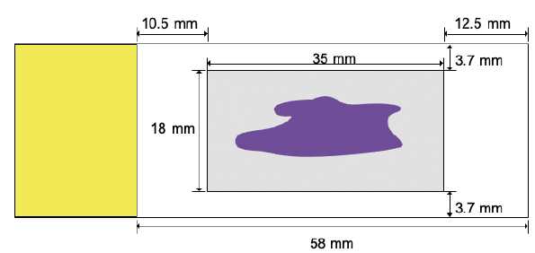
Place a slide in the water bath and gently lay the tissue on the slide as it is removed from the water bath. Make sure the tissue section is placed in the accessible imaging area (gray in image below) of the slide.
Place the slide on an angled slide holder and let it dry overnight at room temperature.
Tissue Pre-treatment
Bake sample slide/s in an incubator at 65°C for 1-3 hours until paraffin thoroughly melts.
Cool sample slide/s at room temperature for 5 minutes to allow the sample slide/s to cool to room temperature.
Tissue Deparaffinization and Hydration
Immerse sample slide/s in a coplin jar containing the following reagents for 5 minutes each:
a.Histochoice
b.Histochoice
c.100% Ethanol
d.100% Ethanol
e.90% Ethanol
f.70% Ethanol
g.50% Ethanol
h.30% Ethanol
i.ddH2O
j.ddH2O
Antigen Retrieval
In a coplin jar, prepare 50 mls of a 1x citrate buffer. Dilute 10x citrate buffer pH6.0 to 1X citrate buffer in ddH2O.
Immerse sample slide/s in the coplin jar containing the 1x Citrate buffer and cover the coplin jar with aluminum foil.
Fill the pressure cooker with 1200 mls of ddH2O. Place the aluminum foil covered coplin jar containing sample slide/s in the pressure cooker.
Set the pressure cooker to the high-pressure protocol and let the tissue incubate for 20 minutes.
After incubation in the pressure cooker, release the pressure and carefully remove the coplin jar from the pressure cooker and allow to cool at room temperature for 30 minutes.
Wash Tissue
Place sample slide/s in a coplin jar containing 50 mls of ddH2O for 2 minutes. All volumes for coplin jar incubations will be 50 mls henceforth in this protocol.
Place sample slide/s in a second coplin jar filled with ddH2O and incubate for 2 minutes.
Place sample slide/s in a third coplin jar filled with 1X PBS and incubate for 2 minutes.
Photobleach Tissue
Submerge the sample slide/s in a 150 mm petri dish containing 50 mls of 4.5% (w/v) H2O2 and 20mM NaOH in PBS (bleaching solution).
Sandwich the petri dish between two broad-spectrum LED light sources for 45 minutes at 4°C.
After 45 minutes move the sample coverslip(s) into a new petri dish with fresh bleaching solution and photobleach for another 45 minutes at 4°C.
Wash Tissue
Wash the sample slide/s three times for 2 minutes each in coplin jars containing 1X PBS.
Immerse the sample slide/s in a coplin jar containing 50 mls of CODEX® Hydration Buffer and incubate for 2 minutes.
Move sample slide/s to a second coplin jar containing CODEX® Hydration Buffer and incubate for another 2 minutes.
Equilibrate Tissue in Staining Buffer
Move sample sample slide/s to a coplin jar containing 50 mls of CODEX® Staining Buffer and incubate for 20-30 minutes.
Preparation of the Antibody Cocktail Solution
Prepare a stock solution of CODEX® Blocking Buffer to be used for the Antibody Cocktail.
| A | B |
|---|---|
| CODEX® Blocking Buffer | 1 Sample |
| Staining Buffer [µL] | 181 |
| N Blocker [µL] | 4.75 |
| G2 Blocker [µL] | 4.75 |
| J Blocker [µL] | 4.75 |
| S Blocker [µL] | 4.75 |
| Total [µL] | 200 |
Add CODEX® Blocking Buffer to a tube designated for Antibody Cocktail Staining Solution. The volume of CODEX® Blocking Buffer to be prepared for each sample slide/s can vary depending on the titer and corresponding volume of each antibody. Adjust volume of CODEX® Blocking Buffer so that the final volume of the Antibody Cocktail Staining Solution is a total of 200 μL per tissue.
Add the appropriate volume of each CODEX antibody to the Antibody Cocktail Solution.
Pipette to mix and briefly spin down the tube.
Tissue Staining
Remove sample slide/s from the coplin jar containing Staining Buffer and place it on the tray of a humidity chamber.
Add 195 μL of the Antibody Cocktail to each of the sample slide(s) and ensure that the liquid covers the entire tissue.
Incubate overnight in the humidity chamber at 4°C.
After overnight antibody incubation, immerse the sample slide/s in a coplin jar containing 50 mls of CODEX® Staining Buffer for 2 minutes.
Place sample slide/s in a second coplin jar containing CODEX® Staining Buffer for 2 minutes.
Fix Tissue
Place sample slide/s in a coplin jar containing 50 mls of 1.6% PFA in CODEX® Storage Buffer and incubate for 10 minutes.
Transfer sample slide/s to a coplin jar containing 50 mls of 1X PBS. Lift and immerse the sample slide/s 2-3 times.
Transfer sample slide/s to a second coplin jar of 1X PBS and immerse sample slide/s 2-3 times.
Transfer sample slide/s to a third coplin jar of 1X PBS and immerse sample slide/s 2-3 times.
Ice-cold Methanol Incubation
Transfer sample slide/s to a coplin jar containing Ice cold methanol and incubate for 5 minutes on ice.
Place a coplin jar of 1x PBS next to the methanol coplin jar containing the sample slide/s. Quickly transfer the sample slide/s from methanol to the coplin jar of 1x PBS and immerse sample slide/s 2-3 times.
Transfer the sample slide/s to a second coplin jar of 1x PBS and immerse sample slide/s 2-3 times.
Transfer the sample coverslip to a third coplin jar of 1x PBS and immerse sample slide/s 2-3 times.
Prepare the Final Fixative Solution by diluting 20 μl of the CODEX® Fixative Reagent in 1 ml of 1x PBS (Final Fixative Solution).
Remove sample slide/s from the coplin jar and place it on the tray of a humidity chamber.
Add 200 μL of Final Fixative Solution to each of the sample slide/s and ensure that the entire tissue is covered in fixative solution.
Incubate for 20 mins.
Remove the sample slide/s from the humidity chamber and place in a coplin jar containing 1x PBS. Lift and immerse the sample slide/s 2-3 times.
Move the sample slide/s to a second coplin jar containing 1x PBS and immerse sample slide/s 2-3 times.
Move the sample slide/s to a third coplin jar containing 1x PBS and immerse sample slide/s 2-3 times.
Store Tissue
Transfer sample slide/s to a coplin jar containing 50 mls of CODEX® Storage Buffer and store at 4°C until imaging.
