ONT DNA Extraction for Fungal Barcoding
Stephen Douglas Russell
Abstract
Below you will find a simple and fast two-step DNA extraction protocol that works well with many different groups of fungi. This protocol can be utilzed in preparation for DNA barcoding of fungi utilizing Oxford Nanopore MinION.
Time required:
Varies depending on the use case. My samples generally have the metadata and images stored on iNaturalist. For each sample, I go to the iNaturalist observation, validate that the specimen matches the metadata and images, include a unique lab code in the "Voucher Number(s)" observational field, record the lab code on the sample packet with a marker, and record the lab code on the associated spreadsheet in the correct cell number.
If all of these actions need to be performed, it can take 3-5 hours per 96 samples to load.
If the iNaturalist observations have already been validated, the lab numbers have been assigned, and all that is required is to put a small amount of tissue in each cell for each sample, then the time outlay can be less than 2 hours per 96 samples.
Steps
X-Amp Extractions
Place 15µL to 25µL of 20µL. Using less solution means the overall cost basis per sample will be lower.
96 well plates could also be used for this extraction protocol. Even though it costs more, I prefer to use 8-strips as they are easier to work with. Only eight sample cells need to be open at a time, individual rows can be removed for closer examination, spin-down at the end is easier, etc.
As you load tissue in each tube, record the information about the sample into a spreadsheet. I use the sheet linked below. Loading each strip from the right to the left will allow the flow from A -> H of a typical 96 well plate style format. I also like to print out the sheets and record them with a pen and pencil so there is a physical record.
Label the plate name with the name of the forward primer being utilized for the plate. Ex - ONT001
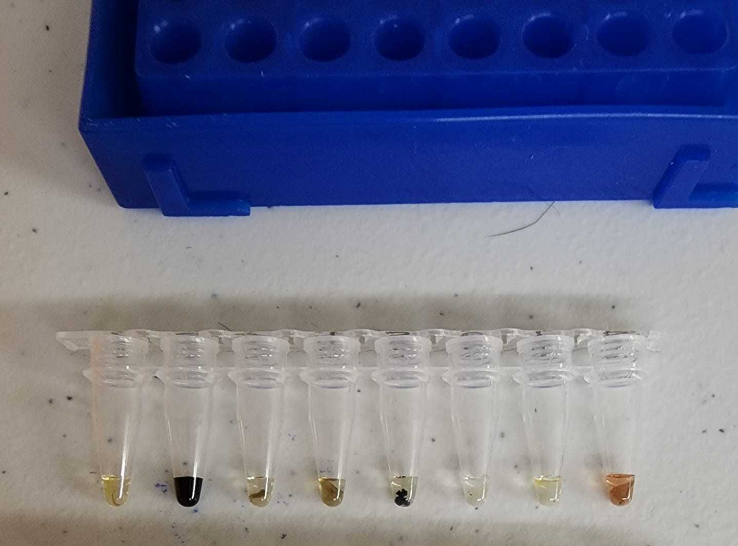
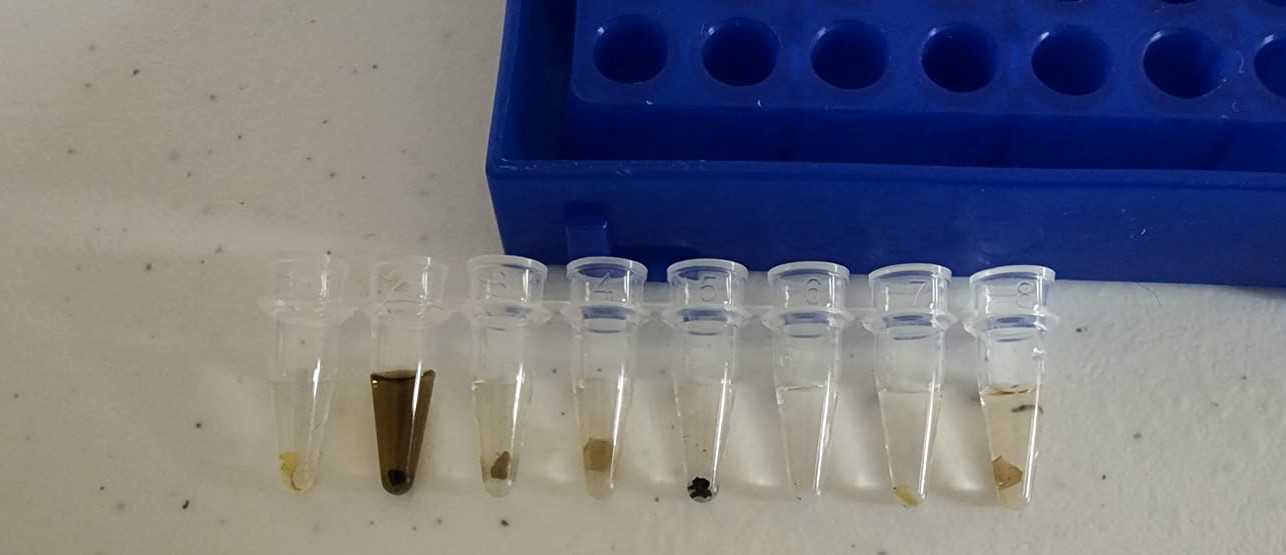
Add a small amount of fungal tissue to each cell. The amount of fungal tissue loaded into each cell should easily be submerged in the amount of extraction solution used (see image). Less is better than more. If you can physically see the sample in the tube it is probably enough for a successful extraction.
For dried specimens, I will typically break off a small section of the mushroom with my fingers and place it on a Kimwipe. From there it can be further broken up on the Kimwipe until you spot a piece of the desired size to put in the tube.
For fresh specimens, I will try to pull off the desired amount for each cell directly from the specimen.
Both dried and specimens use the same visual amount of tissue.
Between specimens, the tweezers and/or teasing needle is wiped off with an alcohol swab and then wiped off with a Kimwipe. I change the alcohol swab and Kimwipe maybe once every one or two 8-strips.

Spin down each strip for 10 seconds in a mini centrifuge. Ensure the tissue in each cell is in the fluid. It is ok if a sample is too large for the amount of fluid and only partially submerged.
Heat the tissue for 0h 15m 0s at 80°C in a thermocycler.


