Handling and Sampling Small Nonhuman Primates - ISL Peru
Gideon Erkenswick, Mrinalini Watsa, Cristian Tirapelle, Alice Poirier, Alexandra Sacco, Priscila Peralta-Aguilar, Efstathia Robakis, Ishaan Raghunandan
Disclaimer
This protocol is actively used by Field Projects International at the Estación Biológico Los Amigos, Madre de Dios, Peru. It is revised annually to reflect improved capture, handling, marking, and sampling methodology. It has been reviewed by the ethics committees of multiple institutions. No author nor affiliated institution takes responsibility or bears any liability for the use of this protocol by others. The protocol is listed as having sensitive content since it involves biosampling from wildlife. Note: these procedures should be carried out only by trained personnel, and are not recommended for use without first obtaining all required permissions.
Abstract
Program Timing:
Trap habituation begins in May. Trapping and sample collection occur annually during the rainforest dry season (June - August). Sample transport and analyses occur between September and April.
Capture Overview:
Entire primate social groups are captured together in multi-compartment traps in the morning to ensure same day release (~ 5 - 30 minutes depending on group sizes that range between 3 and 8 animals) -> animals are administered a small dose of anesthetic for trap extraction and 1st phase processing, then transferred to quiet, dark holding cages until 2nd phase processing (maximum holding cage time of 2 hrs for largest groups) -> one by one, animals are chemically restrained once more for 2nd phase processing for morphometric measurements, photographs, sample collection, and placement of animal identification (~ 25 minutes) -> animals recover in quiet, dark recovery cages during which 3-minute checks for breathing and movement occur until fully alert (1.5-2 h/animal since last injection of anesthetic) -> when the group is fully recovered from anesthetic all are released at the site of capture (< 5 minutes) -> group membership and well-being are confirmed with radio telemetry.
Team Composition:
This protocol is intended to be carried out by a team of no less than 4 individuals, including at least 2 experienced personnel. Roles include (1) veterinarian/senior researcher (2) designated animal handler (3) sampling assistant (4) data recorder. Refer to the PROCESSING ROLES section.
Before start
Trapping events are never spontaneous. They are always planned after careful review of trap habituation data collected during the weeks leading up to trapping. The decision to capture a group is based on observation (either direct or via camera trap footage) of entire social groups reliably entering and feeding inside traps to ensure successful capture of the entire group. The PI is in charge of coordinating the primate capture activities and reviewing habituation data to determine when trapping events take place and at which trap sites.
Prior to trapping, all team members have received prior training on how to process these primate taxa. Typically, all trapping team members participate in several mock trapping sessions beforehand, simulating all steps hereinafter.
The functionality of the trap(s) is checked the day before a trapping event. Primate team personnel pack and double-check that all capture materials needed are cleaned and organized the night before a planned trapping event. Refer to the MATERIALS section.
Steps
Processing roles
Team composition.
This protocol is intended to be carried out by a team of no less than 4 individuals, including at least 2 experienced personnel. Roles include (1) a veterinarian/ senior researcher (2) a designated handler (3) a sampling assistant and (4) a data recorder.
The veterinarian/senior researcher is experienced with every step of the primate capture process. This person is responsible for administering the anesthesia and deciding when an animal might need an additional anesthetic dose and at which point to collect the most invasive samples (biopsy, blood, dental casts).
The designated handler is responsible for handling the anesthetized animal during sample collection, checking vitals to monitor anesthesia (including taking rectal temperature), and disinfecting the sample collection tools in between animals.
The sampling assistant assists with less-invasive sample collection (hair, nails, buccal swabs), taking images listed in the processing sheet, reading aloud the serial codes that identify each sample bag and tube, and passing tools, bags, and sampling tubes to senior researcher(s).
The data recorder records the weight of the animal, morphological measurements, sample numbers, notes from senior researchers, and important timestamps, on the processing sheet. The data recorder also monitors recovering animals every 3 minutes, and stores samples in designated sample boxes as they are being collected. As the team member looking at the processing sheet, the recorder also ensures all relevant samples and information are collected from each animal before completing processing.

Preparatory phase
The day prior to capture, all the materials and equipment necessary for trapping and processing primates are packed and set ready to take to the trap site. The senior researcher decides which trap site will be chosen for the trapping event.
The functionality of the trap(s) is checked the day before a trapping event. Ideally, minimal modifications to trap placement, positioning, and operation are made on the day of capture.
MULTI-COMPARTMENT TRAPS : A string is tied to the outside of each door of the multi-compartment trap (made of galvanized mesh and wood) and passed through a screw eye fixed directly on top of the entrance of each compartment (one for each door). The other end of each string is attached to a tree or rope just in front of a stakeout location where researchers will be seated to monitor the trap. This manual pull system avoids non-target captures and makes it possible to close one door at a time. The strings and trap are setup and tested well in advance. The interior of trap compartments is inspected for sharp points that could cause injury, which are removed or covered as necessary.

On the day of capture, the team arrives at the capture site 30 - 60 minutes before the earliest expected capture time. For these primate taxa, this is before sunrise, as groups may visit a trap first thing upon waking up. This allows for enough time to carry out a final trap inspection, assemble a processing tent, and unpack sampling materials before sunrise.
Two people, at least one being a senior researcher, stay at a designated stakeout location to directly monitor the trap. In some cases, the use of vocalization playbacks may be used to attract the target species to the area sooner, but the decision to use playbacks requires careful consideration of location, time of day, and known identity of the target group.

The remainder of the capture team, usually 2 or 3 personnel , are responsible for assembling the tent and preparing the materials for processing captured animals. The tent is usually set up approximately 50 meters from the trap site, outside of direct visibility from the trap. Following set-up, these personnel remain quiet, out of sight, and ready. They also keep a lookout for any primate movements or calls in the general area in order to give advance notice of target animal(s) approaching the trap.
Communication between the stakeout location and the processing tent is facilitated by walkie-talkies.

Trapping
As soon as the target primate(s) are at the trap, the stakeout team notifies team members in the processing tent via walkie-talkie. All sampling materials are setup on a processing table, and trays and sample collection tools are disinfected. A stopwatch is then started to allow the team to closely monitor the total duration of trapping and processing. A voice recording is also started as a safeguard for ensuring all data are recorded (i.e., morphological measurements, sample tube numbers, etc. that may be missed by the data recorder).
The person starting the recording on the voice recorder reads out the date, the time, the weather conditions, the trap site and primate target group, as well as the team members participating.
For manually capturing primates , a senior researcher positions themselves ready to pull the strings that close the individual trap doors, while the other assists by observing (using binoculars), immediately notifying when a primate fully enters a trap compartment. Animals are trapped one by one in this manner, maintaining tension on the strings so that the trap doors do not fall or get pushed open.

For group capture, the stakeout team waits until all the animals of the group have entered the trap. Ideally, all individuals are trapped within 20-30 minutes or less. Limiting this trapping period reduces stress and risk of injury to animals.
If one or more animals are not entering the trap, a decision is made whether it is safe to leave the animal outside on its own for the duration of processing. For example, if a juvenile is not entering the trap, the team will either release one of the other adult animals or a trapping event may be aborted. In this case, simply releasing tension on the strings allows the animals to exit the trap by themselves. Capture can then be attempted again at a later date.
For single animal captures, once the trap door is shut, the team allows the animal to finish eating the bait while checking to make sure that no other conspecifics are watching before approaching the trap. A single animal may be captured if it is a lone dispersing individual.
Once all group members are believed to have been trapped, the stakeout team scans the surrounding area to ensure no straggling animals are around and watching before approaching to lower the trap. While wearing a face mask, the observer who is not holding the strings quickly approaches the trap and secures all trap doors closed, either using a gloved hand, a rope, or string to hold the front doors closed, as needed. The person holding the strings maintains tension until all doors are secured, and their partner has indicated that they have stabilized the trap. If not stabilized, the trap may tip or fall due to the release of tension.
The person holding the strings then approaches the trap while maintaining string tension, bringing two tarps from the stakeout spot. The long door strings are cut approximately 40 - 60cm from the doors using a pocket knife to avoid the strings getting tangled in the vegetation when the trap is moved to the processing tent. While ensuring the doors remain secured and the trap is stabilized, one person unties the rope tethering the trap to the platform. The trapped animals usually stay in the back of their compartment, away from the person holding the doors.
The trap is then swiftly lowered down to the ground on top of a tarp, with doors facing upwards. The other tarp is used to cover the top of the trap, preventing animals from seeing outside. The tarps ensure that the animals remain calm, and prevent fecal samples from dropping to the forest floor as the trap is transported closer to the processing tent.

1st Phase - Processing
The veterinarian or senior researcher loads syringes with a fixed amount of anesthetic for each animal. Syringes are pre-labeled with the animal capture number and all contain the same volume of anesthetic (around 3 times the estimated volume for an adult individual of that species). Pre-labeling syringes 1- n in accordance with the animal processing order eliminates any confusion about which syringe belongs to which animal.

A veterinarian or senior researcher injects anesthetic through the mesh trap intramuscularly, while the animal is physically restrained with a towel and a leather glove by another team member. Animals are retrained and injected with the anesthetic one at a time.
To restrain an animal, a senior researcher opens the compartment door just enough to quickly slip in a towel while the animal is on the bottom of the trap. While the animal is physically blinded or distracted by the towel, the restrainer pushes the animal toward the side of the trap using a gloved arm. While the restrainer holds the animal in place, the other veterinarian or senior researcher maneuvers into position to inject the anesthetic.


After injection, the team allows the animal approximately two minutes in the covered trap to allow for the anesthetic to take effect. The veterinarian or senior researcher periodically glances at the animal to ensure there are no complicating drug reactions. If an animal is not reacting at all to the first dose after 5 minutes, a second dose may be administered, as it is likely the animal did not receive the full dose during the first attempt.
If the dose was effective, a person calls out the time of pass-out and the animal is then moved into the tent to undergo the first phase of processing. The animal may not be completely immobilized, but the level of sedation is enough to allow for safe handling.

While one person carries the animal into the processing tent, another inspects the empty trap compartment for any feces that might be present. If present, they are collected and placed in a clean, labeled sample bag. The serial code of the sample bag is recorded on the animal’s processing sheet, and the fecal sample is stored in a cooler filled with ice packs.
The tarp placed under the trap also allows for easy retrieval of any feces dropped by the animals, minimizing contamination with the ground.
As soon as the animal is moved inside the processing tent, it is placed inside a cotton bag and weighed using a digital handheld scale. Note: the weight of the empty bag is always recorded by the processing team before placing an animal inside of it.

The animal is then moved onto a clean plastic tray that is disinfected between animals and allows for opportunistic collection of urine or fecal sample collection. A first assessment of the vitals is performed, including rectal temperature, respiratory rate, the color of the mucous membranes and palpebral reflex.
| A | B | C | D | E |
|---|---|---|---|---|
| Time | ||||
| Resp Rate | ||||
| Temperature | ||||
| Color of mucus membrane | ||||
| Palpebral response | Y / N | Y / N | Y / N | Y / N |
The above table is an example of the one found in the primate processing sheet.

If the body temperature is above 101 degrees Fahrenheit, water or alcohol is sprayed on the hands and feet of the animal, and a team member begins fanning the animal to cool its temperature more quickly. Vital assessments are carried out one or two more times during the first phase of processing until temperature and breathing appear normal.
Meanwhile, another person scans the animal to look for previously placed microchip ID tags.

If no microchip is present, the animal is placed in a sternal position and a new microchip is inserted subcutaneously in the area between the shoulder blades.

Once inserted, the microchip number is read out loud, and the corresponding sticker with the identification number is attached to the animal's processing sheet.
1st Phase - Sampling
GLAND SWABS
A small cotton swab is rotated 10 times on the sternal and suprapubic glands and stored in a thermos filled with ice packs, for a microbiome study.


HAIR
A small amount of hair is cut and stored in one sample bag for use in an ecotoxicology project. Additional hair is also plucked from the root and stored in a separate sample bag for use in a DNA project. As a standard rule, all hair is collected from the left flank.

NAILS
The tip of one nail from each hand and foot (4 in total) are collected on a small piece of cotton and stored with the hair DNA sample, in the same sample bag.

VAGINAL SWAB
If an adult female is still under a deeper plane of anesthesia, a vaginal swab is gently rotated in the vaginal canal 2-3 times. The tip of the swab is then cut inside a 1.5mL tube filled with preservative. This sample may be collected during second phase processing if the animal is starting to wake up during the first phase of sampling.
BUCCAL SWABS
Two buccal swabs are collected by inserting a small plastic swab in the buccal cavity and rotating it approx. 5 times. The tip of the swab is then cut inside a 1.5mL tube filled with preservative.


When the animal is awakening or within 15 minutes from the anesthetic injection, the animal is placed inside a cotton bag and moved to an individual compartment in a recovery cage, which is kept inside the processing tent. The cage is covered by sheets to maintain a quiet, dark area for recovery from anesthesia.

Everything that has been in contact with the animal is then cleaned with (1) 10% bleach, (2) water and then (3) alcohol. Gloves are always changed in between animals.


The first phase processing is repeated with each animal.
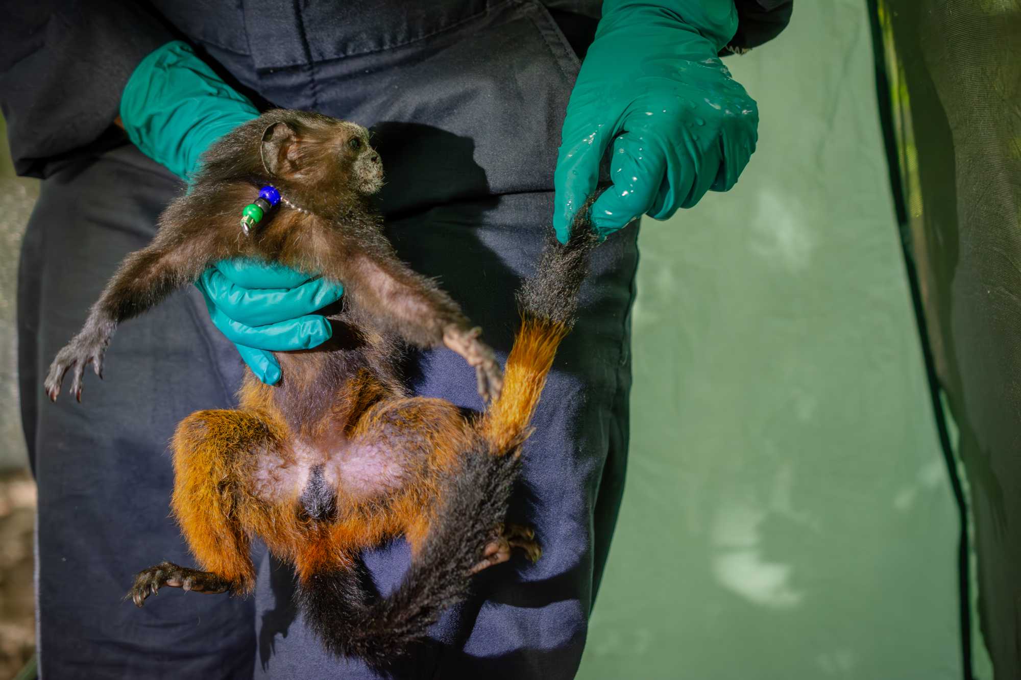
2nd Phase - Processing
The animal, still inside its mesh bag, is removed from the recovery cage, placed on the plastic tray, covered by a light towel, and then injected intramuscularly with the anesthetic, in the thigh. Usually, 2/3 of the total dose (calculated based on the measured body weight of the individual) is enough to allow safe handling of the animal for the second processing.
After about 1-2 minutes, or when induction is achieved, the animal is removed from the mesh bag and vital assessments are recorded (see step #13 ) in the processing sheet.
A team member then measures the tail with a measuring tape before applying a bleach mix to the hair on the tail for a temporary form of identification.
<img src="https://static.yanyin.tech/literature_test/protocol_io_true/protocols.io.5qpvor1exv4o/j2uubx73p.jpg" alt="Image showing different bleaching patterns used for the tail. The letters "M" and "F" refer to the sex of the animal, while the number refers to the individual pattern." loading="lazy" title="Image showing different bleaching patterns used for the tail. The letters "M" and "F" refer to the sex of the animal, while the number refers to the individual pattern."/>
To bleach the tail, the hair-bleaching powder is mixed with liquid activator. The mixture is then applied to the fur on the tail in a specific pattern and covered with aluminum foil. These foils are then left in place for at least 10 minutes to allow decoloration.

2nd Phase - Sampling
Once the vitals are assessed and the bleach is in place, second phase sampling proceeds as follows:
BIOPSY
- The animal is placed on its dorsum, while another team member is covering its head with a light towel.
- Using an antiseptic wipe, the bald skin on the armpit is disinfected.
- Using forceps, a small piece of skin (~ 0.3 mm) is isolated and cut around the tip using fine scissors. The 0.3 mm diameter skin biopsy is then placed in a 1.5mL tube filled with preservative (ideally not a lysis solution).
- A drop of liquid bandage is immediately placed on biopsy wound and allowed to dry.
DENTAL MOLD
-
A new tip is placed on the dental gun and filled with dental impression paste by pressing repeatedly on the gun's trigger.
-
The designated handler holds the animal with one hand, from underneath the armpits.
-
A senior researcher holds the mouth open by holding the top jaw with one hand and holding the thumb of the other hand on the lower incisors, gently pulling down, keeping the animal's mouth open in a stable position.

- The other experienced team member then uses the dental gun to place the dental paste in a "U" shape layer covering the occlusal aspects of the teeth of the lower jaw.

-
A square of laminated paper shaped to fit inside the mouth of the animal is then placed on top of
the first layer of the dental paste. This allows for the upper and lower jaw dental molds to be
collected simultaneously and then easily be separated for later analysis.
-
The entire surface of the laminated square (facing the palate) is covered with dental paste to get
obtain a dental mold of the upper jaw.

-
The animal's mouth is then shut closed and kept stable until the resin solidifies, which takes
about one minute.
-
Once hardened, the dental mold is ready to be removed and stored inside a dedicated sample
bag.
BLOOD
- The designated handler holds the animal with one hand, from underneath the armpits.
- The veterinarian or senior researcher responsible for drawing blood holds the leg of the animal from the knee area (using the non-dominant hand), and holds the syringe with the dominant hand.
- After disinfecting the skin in the femoral area, it is possible to visualize/palpate the femoral artery which is pulsating. To minimize bleeding following venipuncture, we opt to avoid the artery and collect blood from the vein. Parallel to the skin, a 23G needle on a syringe enters around 0.5 cm caudal to the vein, pointing towards the vein at a 90 degrees angle with its longitudinal axis.
- Once the needle enters the blood vessel, there is typically a "flash'" of blood that appears in the plastic neck of the needle. Maintaining this position, the plunger of the syringe is gently pulled back until ~0.5 mL of blood has been drawn.
- After the syringe is carefully removed, the person holding the animal puts strong pressure on the venipuncture site with their thumb to create hemostasis.
- A drop of blood is used for blood glucose and ketone concentration measurements, as well as 2 blood smear slides. A 0.3mL aliquot of blood is also placed in an tube containing lysis buffer. The remainder of the sample is placed in capillary tubes.

2nd Phase - Measurements and Collar placement
Morphometric measurements are then collected using a digital caliper and a measuring tape. All measurements collected are listed in the table from the animal processing sheet below.


Using a camera calibrated with a color card, photographs are also taken of body parts listed in the table found on the processing sheet.


RADIO COLLAR PLACEMENT
If a tracking collar will be placed, its proper functioning is confirmed the day before capture. The collar is placed around the neck, ensuring that it is not putting too much pressure on the throat (too tight) and that if the animal was to try to put its arm through the collar, the elbow cannot get stuck in it (too loose).

BEADED COLLAR PLACEMENT
A beaded collar will be placed around the neck of other group members to allow for individual identification on behavioral follows. Beaded collars are made of rust-resistant aluminum ball-chain string that naturally wears and breaks within 6-12 months; a smaller and weaker version of the material used for army identification tags. Pliers are used to close the aluminum clasp that connects the two ends of the bead chain forming the collar.


The fur of the tail is washed with non-irritating baby shampoo to remove any residual bleach material. The tail is dried with a towel and a full-body picture that includes the bleached tail pattern is taken.

The animal is then left to recover from the anesthetic in a covered recovery cage with individual compartments for each animal.

CLEANING
Clean the surfaces that have been in contact with the animal or its secretions by:
- Spraying 10% bleach and then alcohol;
- Disinfect all the tools used during the processing with the sterilization solutions, accordingly to the protocol: leave submerged in 10% bleach for 3 minutes -> 1st rinse in distilled water -> 2nd rinse in distilled water -> submerge in 70% ethanol.
- Dry all tools and surfaces with clean paper towels or sterile cotton.
The second phase processing is then carried out for the next animal, following the same order used in the first processing
Recovery & Finalization
When all the captured animals have gone through the second processing, they are left to recover inside the recovery cages for about 1.5 h from the time the last injection of anesthetic was received by the last animal processed.
Most of the team members help sterilize equipment and materials, clean the tent, organize samples, and pack up most of the equipment for transportation back to the field station. The voice recording is ended after repeating the following: full date, location, primate group processed, and the names of each team member present.
While most of the team departs the trap site, a senior researcher stays in the tent with the animals until the time of the release: checking them every 10 minutes, making sure they are reactive to stimuli and recovering well from the sedation.
Fecal and urine samples are collected opportunistically from the trays placed underneath each individual compartment of the recovery cage. They are stored in sterile tubes and bags and placed in a thermos container filled with ice packs. Sample numbers are recorded on a data sheet.
Before the release, all the animals are offered banana pieces through the mesh of the recovery cage, which, along with movement and vocalization, helps to assess their readiness for the release and provides them with sugar and hydration.
Release
At least two people must be present and help record the release event. Usually one member of the trapping team comes back to the site at release time, or a different researcher joins the senior researcher.
The recovery cage is carefully moved outside of the tent, still covered with the sheet, and placed on the ground in a suitable release spot. The senior researcher kneels down near the cage, ready to open the compartments, while the second person is positioned five meters behind, holding binoculars or a camera, ready to observe the release. A video recording is started to document the release.
Each compartment is opened consecutively so that the animals exit the recovery cage one at the time. This allows taking individual pictures that can be used by researchers to learn to recognize the group members.
The observers remain in the area for a few minutes until they can ensure that all the animals are moving normally. In the case in which one or several animals were left out during during the trapping event, the observers ensure they witness the reunification of these animals with the rest of the group. Usually the animals that are not trapped will remain close by for the duration of the trapping event, often heard long-calling in search of their group mates, and reunite with them soon after release. If group reunification does is not witness after release, the group is followed in days following the trapping event in order to ensure the group is complete.
Sample sorting
After release, the rest of the equipment is packed up and brought back to the station, where sample sorting and data entry then take place.
SAMPLE SORTING
Organize all samples according to the laboratories sample storage protocol.
IMPORTANT: serum samples must be spun and extracted to a serum storage tube the same day and then stored frozen. Do NOT freeze serum tubes until they have been spun down.
If sample sorting is delayed for any reason, samples should be stored in the freezer, unless otherwise indicated by a senior scientist.
FIX BLOOD SMEARS
- Arrange slides face-up on a clean surface and confirm that sample code is clearly visible. If not, trace over to make a clear labeling
- Identify a coplin jar with methanol that has not expired and is full.
- Open the jar and place smears inside (pairs can be placed back-to-back, MAKE SURE THAT SMEAR IS FACING OUTWARD). Quickly close jar again to prevent the methanol from oxidizing.
- Leave the smears in solution for 5 minutes
- Wearing a pair of gloves, remove each slide, gently tap the edge on an absorbent paper to remove the excess solution, and place flat on an absorbent paper to dry for a few minutes. Make sure to close the coplin jar as quickly as possible to preserve to the methanol.
- Place in the storage box.
Data Organization and Entry
For the remainder of the day, the team will:
Check that all the information on the processing sheet has been entered correctly and that the sample numbers match the data, and listen to the voice recording to fill in missing information on the data sheet with a differently colored ink pen (usually red).
Replenish and repack supplies for the next trapping event. Usually part of the trapping team prepares and pre-pack the supplies, then the rest of the team double-checks the packing, to ensure nothing is missing.
Upload the information gathered on the PROCESSING SHEETS to the online ODK form.

Write a detailed narrative report of the capture session. This is the TRAPPING REPORT .
Gather pictures and videos and sort them into designated folders on the project hard drive.
Convert the processing sheets of each individual into PDF files, and save them into each animal's dedicated folder.
Group all the files into a unique folder named by date of capture and by animal number [yyyymmdd_group_animalname].
Save the VOICE RECORDER file into the folder.
Wash all the linens used during the capture (i.e. coveralls, animal bags, towels, sheets used to cover the recovery cage) with detergent and a disinfectant product, such as quaternary ammonium based solutions.
Put all batteries to charge. We are using rechargeable batteries which should preferably be charged during daylight, when the solar panels that provide electricity to the station are working.
