Electroporation transformation of Ostreococcus tauri
Shikha Adhikari, Anbarasu Karthikaichamy
Disclaimer
DISCLAIMER – FOR INFORMATIONAL PURPOSES ONLY; USE AT YOUR OWN RISK
The protocol content here is for informational purposes only and does not constitute legal, medical, clinical, or safety advice, or otherwise; content added to protocols.io is not peer reviewed and may not have undergone a formal approval of any kind. Information presented in this protocol should not substitute for independent professional judgment, advice, diagnosis, or treatment. Any action you take or refrain from taking using or relying upon the information presented here is strictly at your own risk. You agree that neither the Company nor any of the authors, contributors, administrators, or anyone else associated with protocols.io, can be held responsible for your use of the information contained in or linked to this protocol or any of our Sites/Apps and Services.
Abstract
The protocol describes steps for electroporation transformation of Ostreococcus taurii. This protocol is a direct version of published transformation protocol, https://dx.doi.org/10.3791/4074 by van Ooijen et al.
Steps
Preparing Ostreococcus taurii culture
Culture Ostreococcus taurii cells in K-media. The protocol used to prepare K-media is
Sub-culture Ostreococcus taurii cells in K-media at 1% dilution every 10 days and grow in a plant growth chamber under constant light and a Lee Moonlight Blue filter 183 (http://www.leefilters.com/lighting/colour-details.html#183&filter=cf).
Keep light intensity at 20 μmol m-2 s-1 and temperature at 23°C . Shake the every 1 to 3 days to reduce aggregation.
Collect 50mL cells for each transformation at a a cell density of 20-30 x 10^6 mL-1, about 7 days after subculturing. Count approximate cell density using a haemocytometer at 40x magnification.
Electroporation
Prepare DNA by using the Qiagen midi prep kit to obtain 5µg of pure plasmid DNA in a concentration of 1µg/µL in sterile deionized water. Digest product with an appropriate single cutter restriction enzyme to linearize the plasmid. Purify through ethanol precipitation or PCR purification kit. Re-suspend or elute the linearized plasmid in appropriate amount of sterile deionized water.
Prepare microcentrifuge tubes containing 5µg linearized plasmid DNA for each transformation. A control with no DNA is necessary for transformation. Keep these tubes on ice, together with a 2 mm electroporation cuvette for each transformation. The plasmid volume should not exceed 10% (v/v) of the transformation mix.
Prepare 1Molarity (M) solution of Sorbitol in ddH2O and add 0.1% (v/v) pluronic acid F68. Filter sterilize this solution. Prepare 2.2 ml of resuspension buffer for each transformation.
Add pluronic acid F68 to the cells up to a final concentration of 0.1% (v/v). Centrifuge the cells for0h 10m 0s at 8000x g,10°C,0h 0m 0s in a 50mL tube. Pipette up and down to re-suspend the cells in 1mL solution of resuspension buffer and transfer to a microcentrifuge tube. Centrifuge the tube for 0h 10m 0s at 8000x g,10°C,0h 0m 0s. Repeat this wash.
Resuspend each final pellet in 40µL of resuspension buffer. After resuspension, add 40µL of cells to each tube of linearized DNA. Keep linearized plasmid on ice while mixing gently with a pipette, and transfer to an electroporation cuvette.
Use a cut tip or 1000µL tip to avoid damaging the cells.
Electroporate the cells using the following electroporation parameters,
Equipment
| Value | Label |
|---|---|
| Gene Pulser Xcell Electroporation Systems | NAME |
| Electroporator | TYPE |
| Biorad | BRAND |
| 1652660 | SKU |
Voltage: 6 kV cm-1 (since we are using 2 mm cuvette, the voltage setting would be 1200 V)
Resistance: 600 Ω
Capacitance: 25 μF
Incubate cells in cuvettes at -90Room temperature for 0h 10m 0s. Prepare T25 tissue culture flasks by adding 30mL of fresh K-media and labeling each. Take 1mL of K-media out of a flask and add it to the corresponding cuvette, making sure to pipette up and down carefully to move the globule of cells into the correct flask. Make sure not to disturb the cell globule.
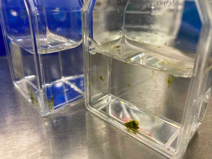
Place cells in the plant growth chamber for 1h 0m 0s to 2h 0m 0s to allow them to recover. Re-suspend by shaking flasks, making sure no clumps are visible. Let cultures recover overnight in the growth chamber.
Inclusion of Cells on Plates in Semi-solid Medium
Autoclave 2.1Mass Percent LMP agarose in ddH2O and keep at 65°C to 90°C, under constant stirring. For each transformation reaction, prepare 8x 55 mm diameter petri dishes and 8x 15mL tubes. Each tube should contain 9mL of K-media and 2mg/mL G418.
Take the cells from the growth chamber into a sterile flow hood. Add 1mL of LMP agarose to each of the 15mL tubes with 9mL K-media, close and mix by inverting. Mix 0.5mL of transformed cells and quickly pour it into a 552mL mm diameter plate. Repeat for the remaining tubes.
Allow agarose to set by leaving plates open inside the flow hood for 1h 0m 0s, then close the plates. Place the plates in large square Petri dishes, with four plates per square dish. Parafilm seal the square plates and carefully set the plates in the growth chamber.
Selection of Transformed Colonies
The transformation plates should host colonies within 2 to 3 weeks. Pick up colonies using a 200µL pipette with tips cut off.
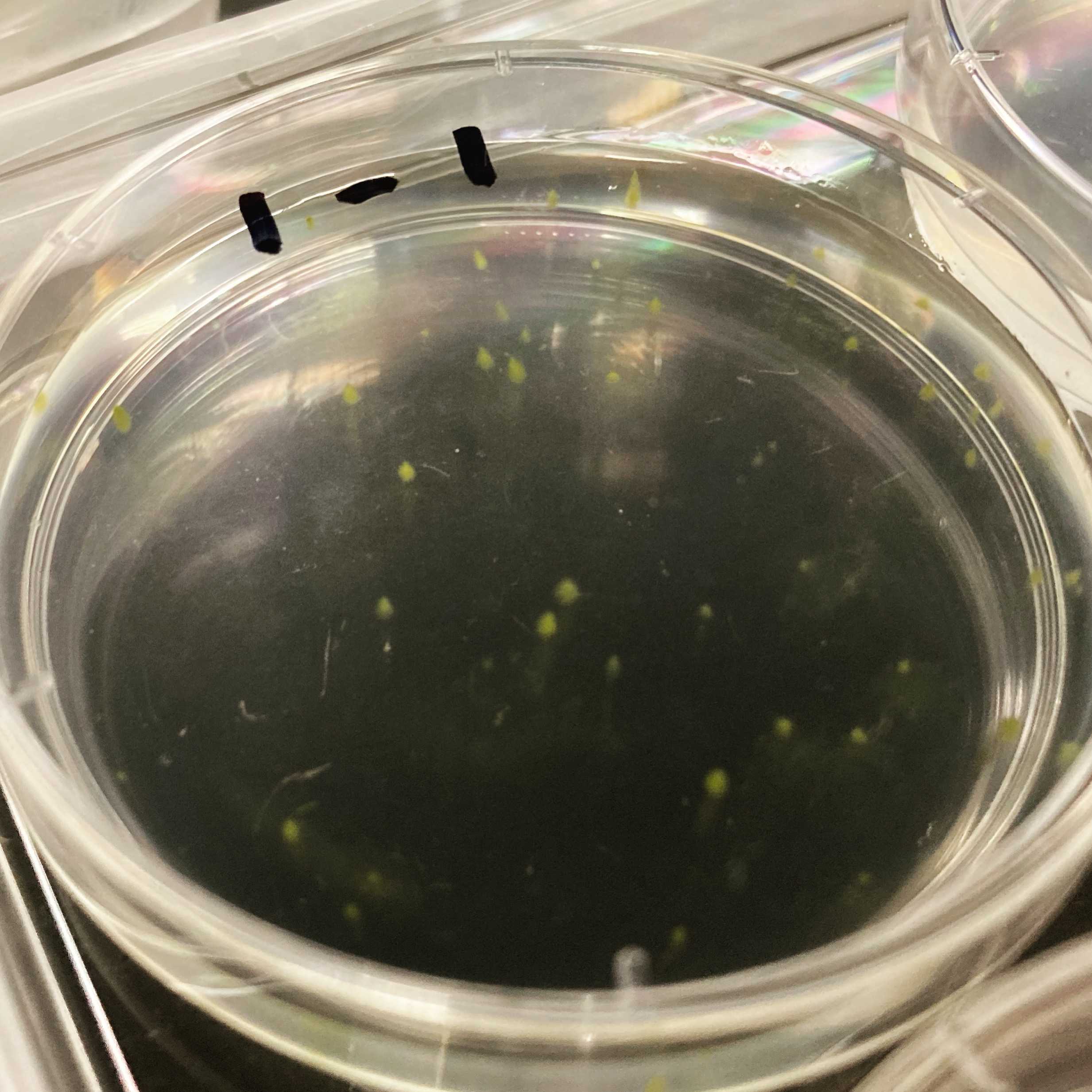
Use a 24 well plate to transfer cell 24-50 colonies from each transformation into 2mL of K-mdeia with 2mg/mL G418 each. Parafilm seal the plates and place in the growth chamber.
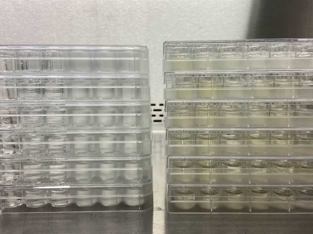
After a week, transfer 100 uL of each well into 2mL of fresh K-mdeia with 2mg/mL G418 into another 24 well plate and grown for a week. Use colony PCR to analyze stable integration of the plasmid DNA.
Transformation efficiency is around 300 CFU/ug plasmid
Representative gel image showing DNA bands for positive Ostreococcus transformants.

