Doubling the resolution of a confocal spinning-disk microscope using image scanning microscopy
Shun Qin, Sebastian Isbaner, Ingo Gregor, Jörg Enderlein
Published: 2020-11-26 DOI: 10.1038/s41596-020-00408-x
Fluorescence Microscopy
Image Scanning Microscopy
Confocal Spinning-Disk Microscope
Structured Illumination
Resolution Doubling
Supplementary information
Supplementary Information
Supplementary Note 1, Supplementary Figs. 1–7 and Supplementary Table 1.
Supplementary Video 1
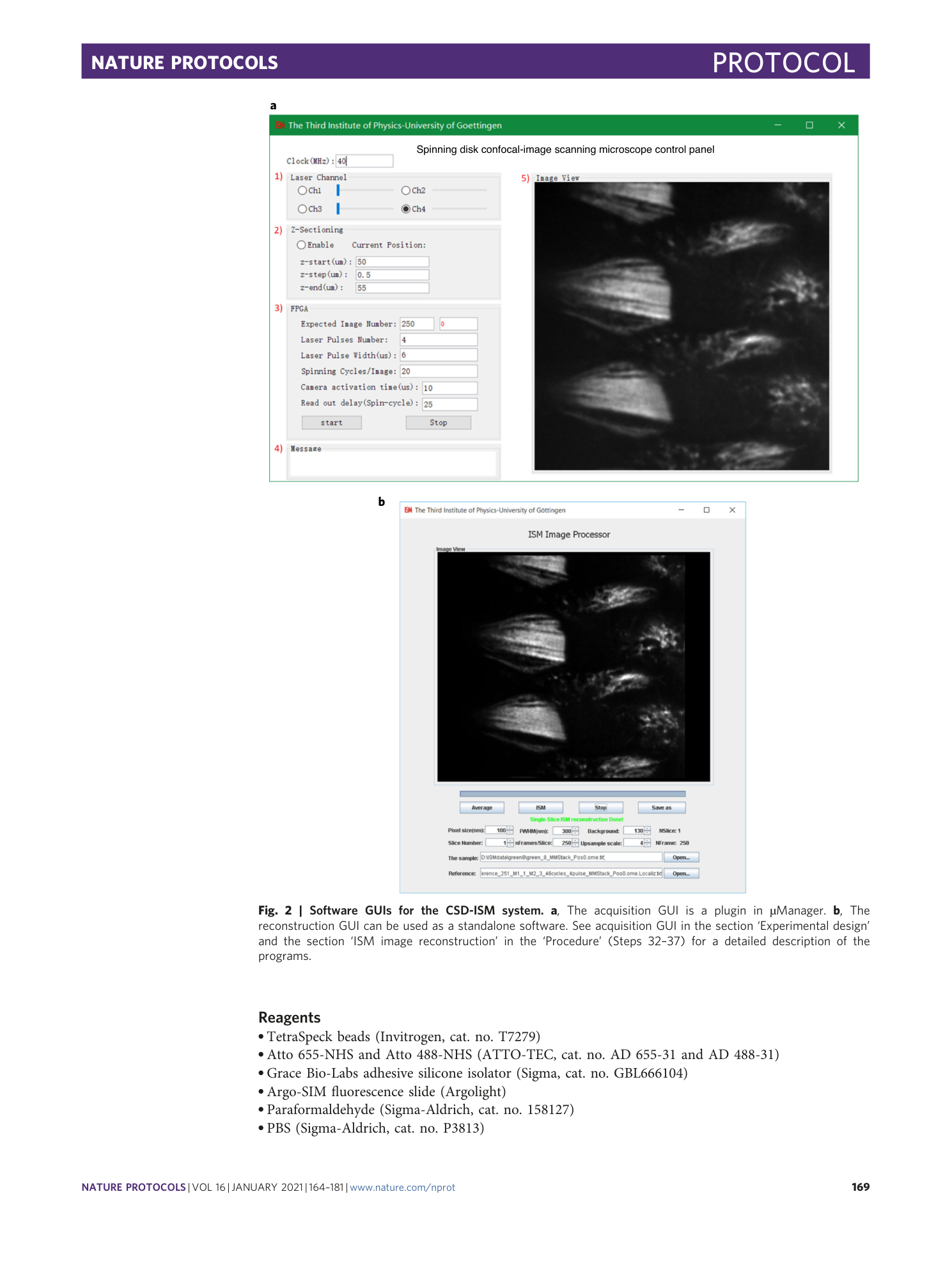
Screen-captured video explaining the use of the Micro-Manager plugin.
Supplementary Video 2
Screen-captured video explaining the use of the reconstruction software.
Supplementary Video 3
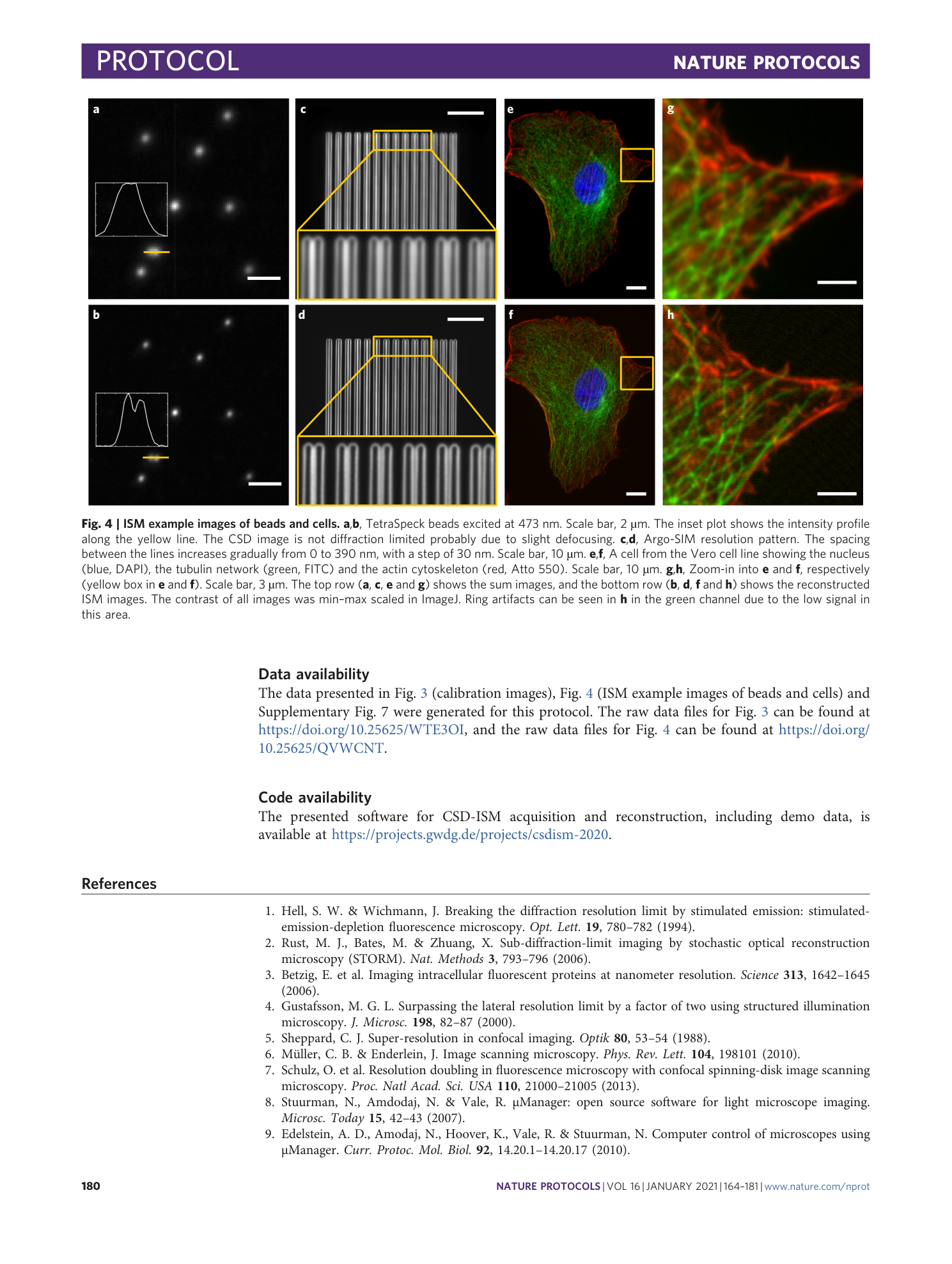
This video illustrates the data acquisition and the ISM reconstruction of the bead image in Fig. 4a,b.
Supplementary Video 4
This video illustrates the data acquisition and the ISM reconstruction of the cell image in Fig. 4g,h.
