ChIP-seq Library preparation

Vasso Makrantoni, Daniel Robertson, Adele L. Marston

Published: 2021-09-22 DOI: 10.17504/protocols.io.bn35mgq6

Abstract

A plethora of biological processes like gene transcription, DNA replication, DNA recombination, and chromosome segregation are mediated through protein–DNA interactions. A powerful method for investigating proteins within a native chromatin environment in the cell is chromatin immunoprecipitation (ChIP). Combined with the recent technological advancement in next generation sequencing, the ChIP assay can map the exact binding sites of a protein of interest across the entire genome. Here we describe a-step-by step protocol for ChIP followed by library preparation for ChIP-seq from yeast cells.

Before start

Steps

ChIP-seq Library preparation

1.

Note
There are commercially available kits for generating DNA libraries but it is relatively straightforward and cost effective to create libraries using standard molecular biology reagents and custom oligonucleotides. This protocol can be completed within 1 day, and it comprises five distinct steps: blunting reaction; dA-Tailing to the 3′ end of the DNA fragments; adapter ligation to the DNA fragments; PCR for enrichment of adapter modified DNA fragments; and library size selection. Finally, given the high cost of ChIP-seq runs and the time-intensive bioinformatics analysis and data validation, it is essential that the quality and the concentration of the libraries is validated by an Agilent Bioanalyzer prior to sequencing.

DNA End-Repair

2.

Perform the blunting reaction using the following recipe: if making multiple libraries prepare a master mix of buffer, dNTPs, and enzyme, and then aliquot to the required ChIP purified DNA. The final volume should be 50 μl.

(a) 1ng20ng (ideally 2 ng)

(b) 5µL

(c) 5µL

(d) 1µL

Note
For library preparation we recommend to use only filter-tips and 1.5 ml DNA LoBind tubes.

3.

Incubate at 65Room temperature for 0h 45m 0s.

4.

Perform a 1.6:1 AMPure XP selection by adding 80µL to the 50µL blunting reaction (see protocol "AMPure Purification Protocol").

5.

Elute in 30µL and take 27.7µL to a new DNA LoBind Eppendorf.

“A”-Tailing Reaction

6.

Use end-repaired DNA (from step 5) to perform “A”-tailing reaction using the following recipe: if making multiple libraries prepare a master mix of buffer, dATP, and enzyme, and then aliquot to the required end-repaired DNA. The final volume should be 30 μl.

(a) 27.7µL

(b) 3.3µL

(c) 1µL

(d) 1µL

7.

Incubate at 37°C for 0h 30m 0s.

8.

Heat-inactivate Klenow enzyme at 75°C for 0h 5m 0s.

9.

Place reaction On ice for 0h 5m 0s.

10.

Proceed immediately to adapter ligation reaction (next step).

Adapter Ligation Reaction

11.

Use dA-tailed DNA (from previous step) to perform the adapter ligation reaction using the following recipe:

the final volume should be 70 μl. Use different barcoded adapters for each Input and IP sample.

(a) 33µL

(b) 35µL

(c) 1µL [15]

(d) 1µL

Note
Always keep adapters on ice. The quantity of adapters stated here is recommended for 2 ng of DNA; however, the amount of adapters should be proportional to the amount of DNA used. Optimal concentration of adapters used is essential. High concentration can lead to adapter contamination in the final library, which can be visualized on the Bioanalyzer (Fig.Always keep adapters on ice. The quantity of adapters stated here is recommended for 2 ng of DNA; however, the amount of adapters should be proportional to the amount of DNA used. Optimal concentration of adapters used is essential. High concentration can lead to adapter contamination in the final library, which can be visualized on the Bioanalyzer (Fig.2b). We use for this protocol the NEXTflex DNA Barcodes—12 (Bioo Scientific; #NOVA-514102). The NEXTflex DNA Barcodes utilize an indexed adapter containing a 6 nt unique sequence. Details can be found in manufacturer’s manual.). We use for this protocol the NEXTflex DNA Barcodes—12 (Bioo Scientific; #NOVA-514102). The NEXTflex DNA Barcodes utilize an indexed adapter containing a 6 nt unique sequence. Details can be found in manufacturer’s manual.

12.

Incubate at Room temperature for 0h 25m 0s.

13.

Perform a 1:1 AMPure selection by adding 70µL to the 70µL (see protocol "AMPure Purification Protocol").

14.

Elute in 52µL. Transfer 50µL to a new DNA LoBind Eppendorf.

Note
DNA fragments over 100 bp will bind to beads and be eluted.

15.

Perform another 1:1 AMPure selection by adding 50µL to the 50µL from the previous step.

16.

Elute in 33µL and take up 30µL to a new DNA LoBind Eppendorf.

Note
DNA fragments above 100/200 bp will bind to beads and be eluted.

PCR Amplification Reaction

17.

Transfer 10µL per sample (previous step) to a 0.2 ml PCR tube and set up the following PCR reaction On ice in a final volume of 50 μl.

(a) 10µL

(b) 10µL

(c) 4µL

(d) 2µL

(e) 1.5µL

(f) 0.5µL

(g) 22µL

18.

Amplify DNA with the following PCR program:

ABC
TemperatureTimeCycles
98 °C30 sec
98 °C10 sec12 -18 cycles
65 °C30 sec
72 °C30 sec
72 °C5 min

Note
When PCR for library amplification is performed, minimal cycling is desirable. The fewer number of PCR cycles used to amplify libraries, the less biased the resulting libraries will be for the products that are more efficiently amplified. Overamplification can result in daisy-chains of fragments that can be visualized as a higher molecular weight peak on the Bioanalyzer. If the library amplification fails, more DNA template can be used. For IP samples use up to 20 μl of template DNA.

Double-Sided AMPure Selection and Library Elution

19.

Perform a 0.65×:1 AMPure selection. To the 50µL (previous step) add 31.85µL, resuspend by pipetting and leave at Room temperature for 0h 10m 0s to bind.

Place 0.2 μl PCR tubes in a DynaMag-PCR magnet for 0h 5m 0s.

20.

KEEP THE SUPERNATANT , this will contain fragments <300 bp. To 80µL add 50µL (adjust bead volume for smaller supernatant volumes) and perform AMPure selection according to steps in protocol "AMPure Purification Protocol".

Note
DNA fragments of 100–250 bp will bind to the beads.

21.

Elute in 52µL and pipet 50µL to a new DNA LoBind Eppendorf.

22.

Perform another 1:1 AMPure selection by adding 50µL to the 50µL from the previous step.

Note
DNA fragments above 100/200 bp will bind to the beads.

23.

Elute in 33µL and transfer 30µL to a new LoBind Eppendorf.

24.

Determine DNA library concentration by Qubit HS kit (use 2µL).

25.

Run the library on a Bioanalyzer to determine average fragment size and general purity. Use a High Sensitivity DNA Kit (Agilent Technologies) as per manufacturer’s instructions. Fragments of the sequencing library should have a size range of 150–300 bp (Fig.2b, upper panel). If not pure, that is, adapter dimers are visible (Fig.2b, lower panel), perform 1:1 AMPure purification to remove small adapter dimers.
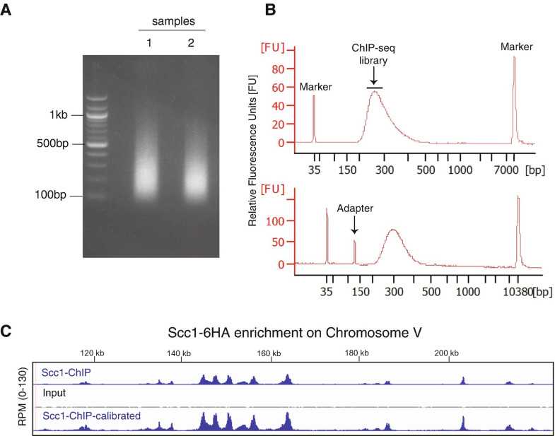
(a) Representative image of mitotic yeast cells sonicated with a Bioruptor Twin (Diagenode) for a 30-min round (power setting: High, 30 s ON/30 s OFF). DNA from two different samples was loaded on a 2% agarose gel with a 100 bp marker ladder. (b) Representative optimal BioAnalyzer trace (upper panel) and contaminated trace with adapter (bottom panel) (c). Examples of profiles generated by chromatin immunoprecipitation followed by sequencing (ChIP–seq) of the cohesin subunit Scc1 in wild-type cells (IP shown in blue; Input shown in grey) and calibrated with S. pombe Scc1 distribution in representative chromosome V (IP shown in blue, bottom panel)
26.

Libraries are now ready for sequencing using a sequencing platform of choice.

27.

The final concentration of the library to load on the flow cell is 1.5 pM with an Input–IP ratio 15%:85%. Perform paired-end sequencing with 76 bp—76 bp for Read 1 and Read 2.

Note
Several different next generation sequencers are available; for this protocol we use the Illumina platform. While platforms vary by target sequence length, accuracy and cost all give reproducibly comparable results. In order to perform experiments in a cost-effective manner, multiplexing can be used, that is, multiple ChIP-seq libraries, each carrying a different barcode to identify different samples can be sequenced together on a single flow cell of MiniSeq or lane of a Hiseq. The output of the Illumina MiniSeq is ~25 M clusters. We typically, sequence 8–10 barcoded uncalibrated samples in a single pooled library or 4–5 barcoded calibrated samples in a single pooled library. Typically one ChIP library generates six to ten million reads [Several different next generation sequencers are available; for this protocol we use the Illumina platform. While platforms vary by target sequence length, accuracy and cost all give reproducibly comparable results. In order to perform experiments in a cost-effective manner, multiplexing can be used, that is, multiple ChIP-seq libraries, each carrying a different barcode to identify different samples can be sequenced together on a single flow cell of MiniSeq or lane of a Hiseq. The output of the Illumina MiniSeq is ~25 M clusters. We typically, sequence 8–10 barcoded uncalibrated samples in a single pooled library or 4–5 barcoded calibrated samples in a single pooled library. Typically one ChIP library generates six to ten million reads [23,24]. However, the above is subject on the level of enrichment of the protein of interest and the resolution required.,Several different next generation sequencers are available; for this protocol we use the Illumina platform. While platforms vary by target sequence length, accuracy and cost all give reproducibly comparable results. In order to perform experiments in a cost-effective manner, multiplexing can be used, that is, multiple ChIP-seq libraries, each carrying a different barcode to identify different samples can be sequenced together on a single flow cell of MiniSeq or lane of a Hiseq. The output of the Illumina MiniSeq is ~25 M clusters. We typically, sequence 8–10 barcoded uncalibrated samples in a single pooled library or 4–5 barcoded calibrated samples in a single pooled library. Typically one ChIP library generates six to ten million reads [23,24]. However, the above is subject on the level of enrichment of the protein of interest and the resolution required.]. However, the above is subject on the level of enrichment of the protein of interest and the resolution required.

推荐阅读

Nature Protocols
Protocols IO
Current Protocols
扫码咨询