AT8 Tau Pathology Image Analysis
Hemanth Ramesh Nelvagal, Toby J Curless, Zane Jaunmuktane
Abstract
QuPath is a bioimage analysis software designed for digital pathology and whole slide image analysis. This protocol describes how to analyse AT8 tau pathology in human brain tissue (FFPE sections with IHC).
Steps
Annotation
Manually annotate regions of interest on NZConnect (Hamamatsu), a web-based whole-slide image (WSI) viewerhttps://www.hamamatsu.com/us/en/product/life-science-and-medical-systems/digital-slide-scanner/U16179-01.htmml.
Download annotations using a Python script.
QuPath De-Convolution and Measurements
Import into QuPath using a Groovy script. Refer to: Bankhead, P., Loughrey, M.B., Fernández, J.A. et al. QuPath: Open source software for digital pathology image analysis. Sci Rep 7 , 16878 (2017). https://doi.org/10.1038/s41598-017-17204-5
In QuPath, apply colour deconvolution to distinguish DAB from the haematoxylin counterstain.
Measure the area of positive DAB staining for tau pathology using a fixed threshold value of 0.2 on the DAB deconvolved channel.
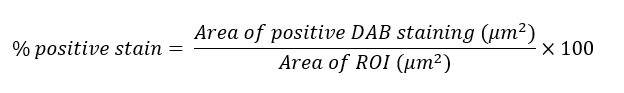
Calculate the percentage of positive DAB staining within the ROI by calculating the area of positive
DAB staining divided by the area of the ROI and multiplied by 100.