Tissue Sectioning Guidelines (FFPE) - CODEX/PhenoCycler
William F Flynn, Elise Courtois, Santhosh Sivajothi, Emily Soja
Abstract
The purpose of this SOP is to provide general guidelines for preparing FFPE tissue sections suitable for PhenoCycler/CODEX
Steps
Tissue Sectioning Guidelines for CODEX/PhenoCycler
Prepare the microtome for sectioning. Section the tissue at a thickness of 5 μm .
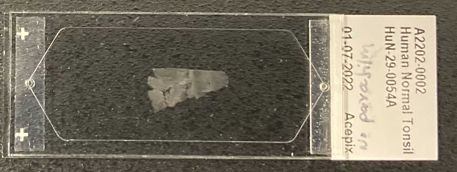
For best results, the tissue should be completely adhered to the slide with minimal tears or folds.
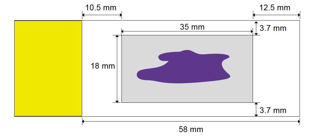
Gently place the tissue section in the center of the slide within the imageable area as shown in
grey rectangle below.
If tissue overlaps the boundary, proper coverslip seal will not be formed. Multiple tissues can be placed within the boundary, as long as there is no overlap of paraffin over adjacent tissues

Dry tissue sections overnight at RT
After drying, store cut slides at 4°C (for up to six months)
NOTE: Do not place any stickers or labels on the frosted end of the slide as it may interfere with coverslip adhesion. Use markers or directly print labels on the slide.


