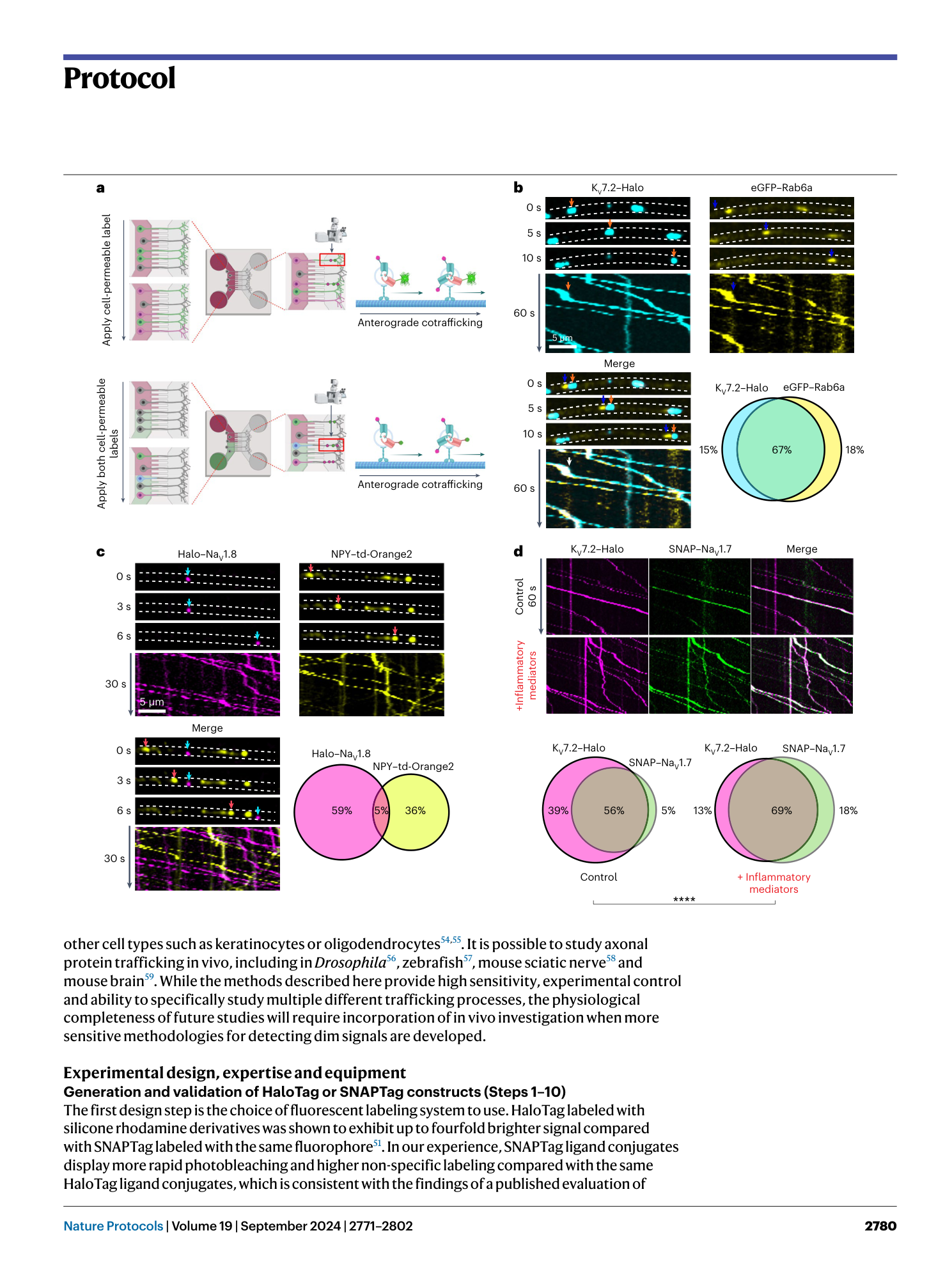
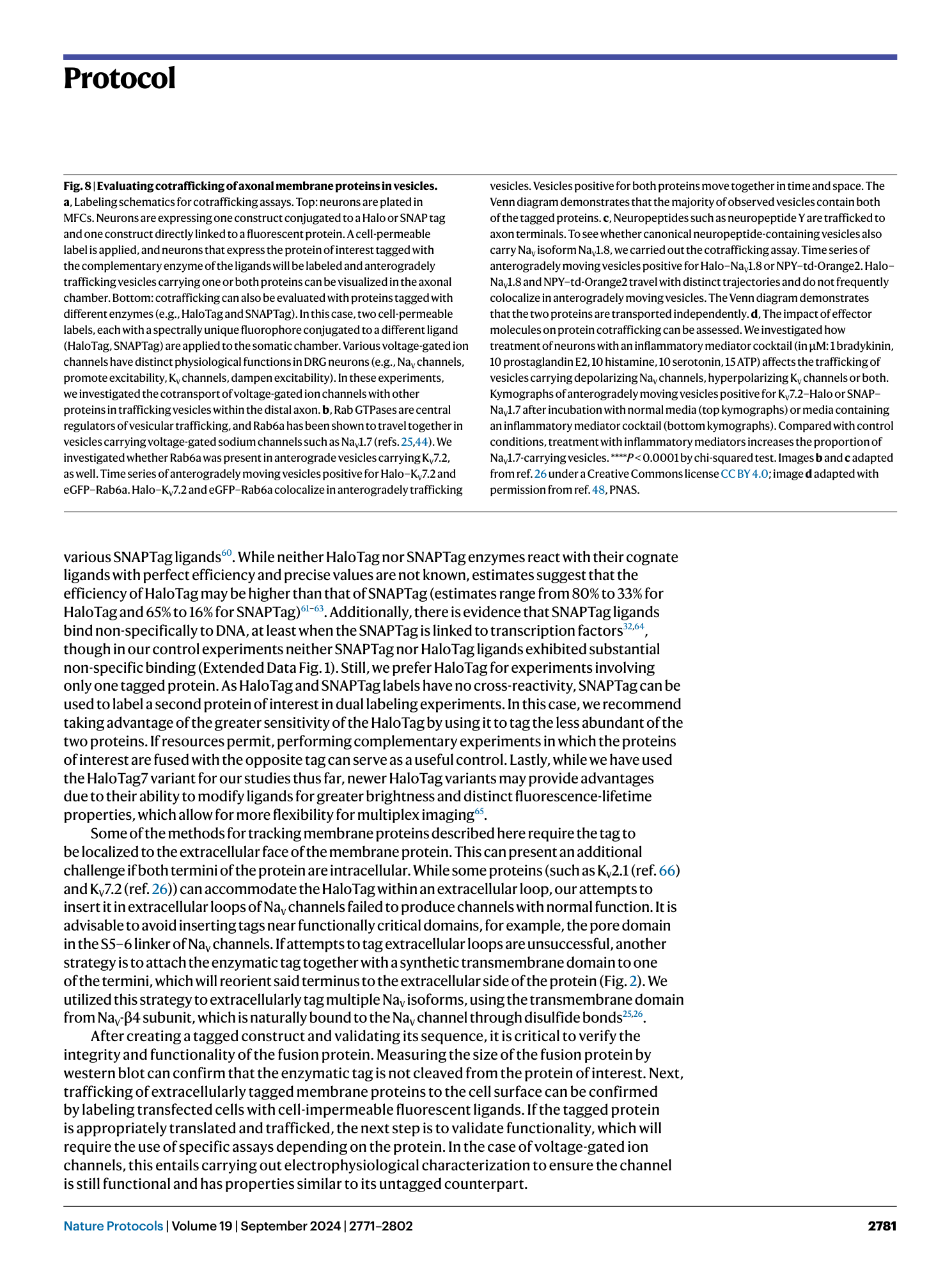
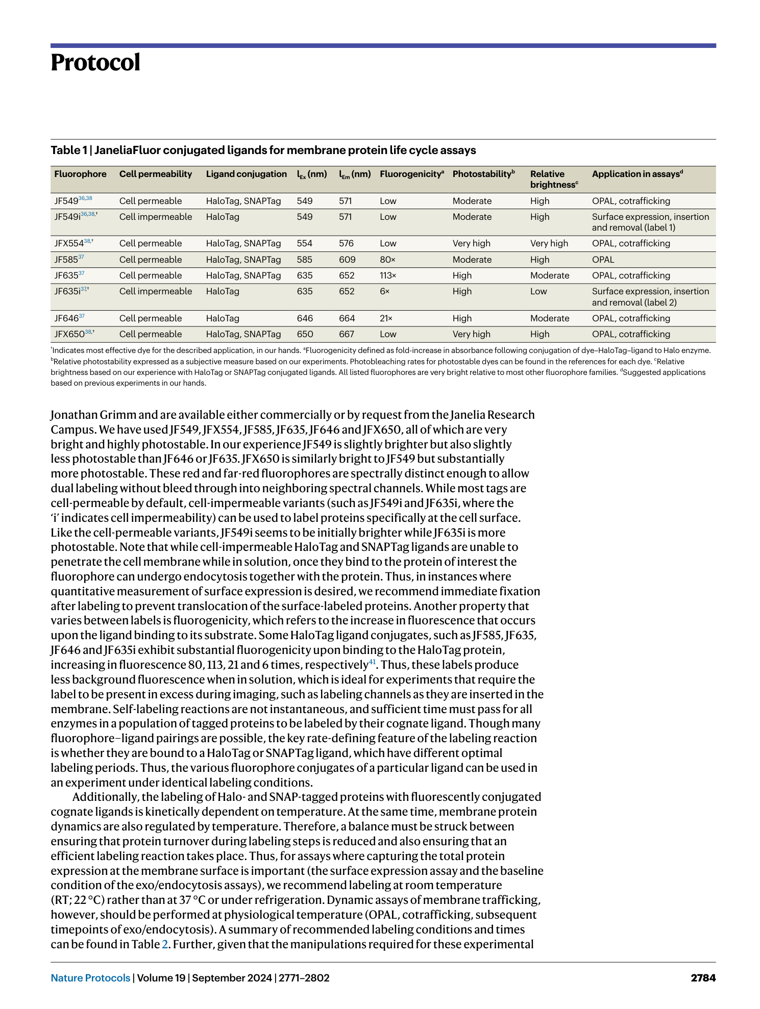
Real-time imaging of axonal membrane protein life cycles
Sidharth Tyagi, Grant P. Higerd-Rusli, Elizabeth J. Akin, Christopher A. Baker, Shujun Liu, Fadia B. Dib-Hajj, Stephen G. Waxman, Sulayman D. Dib-Hajj

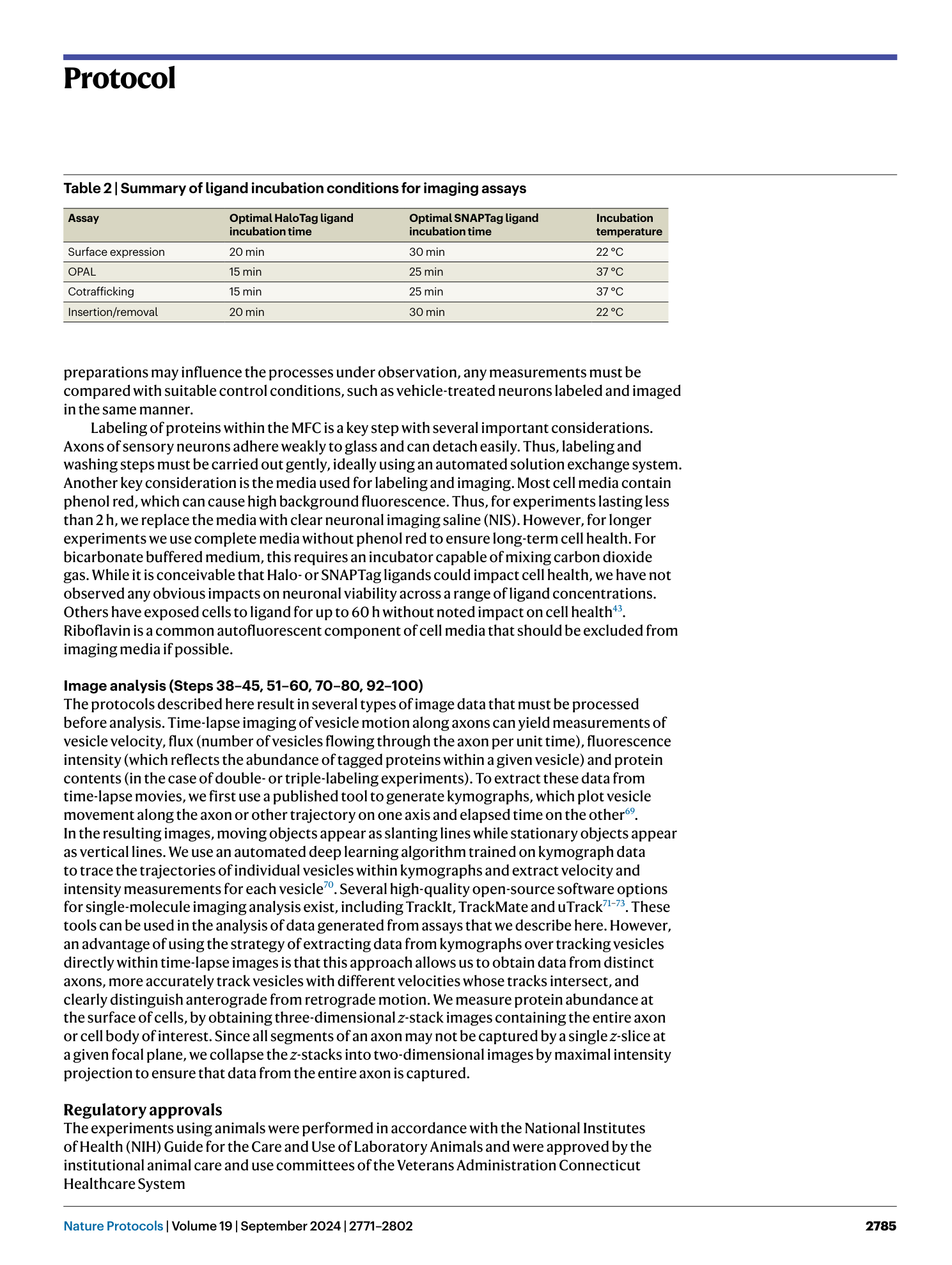


Extended
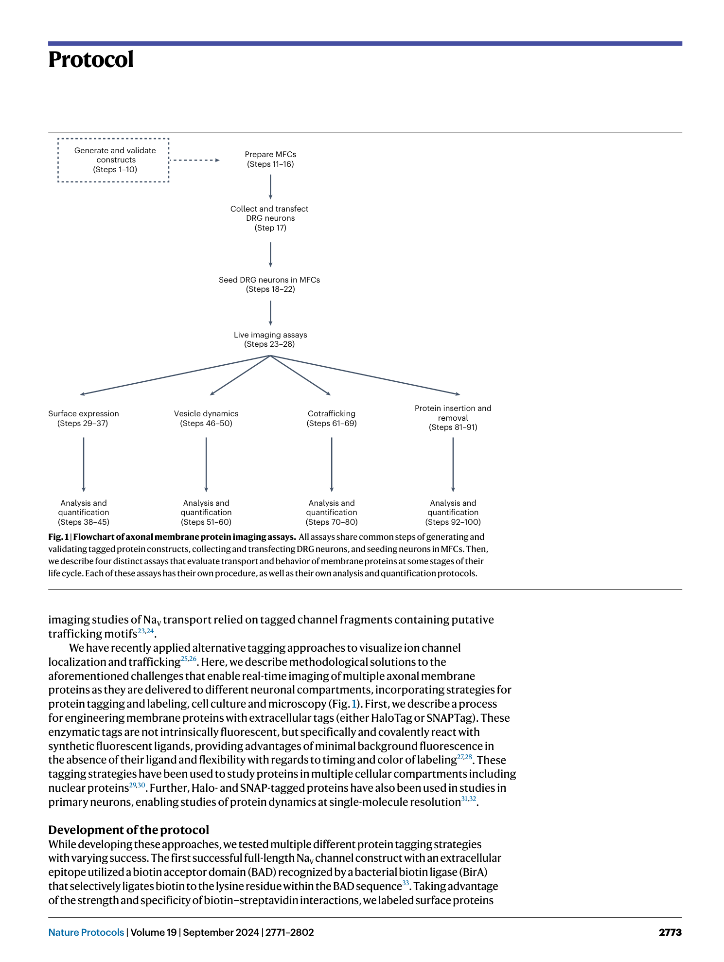
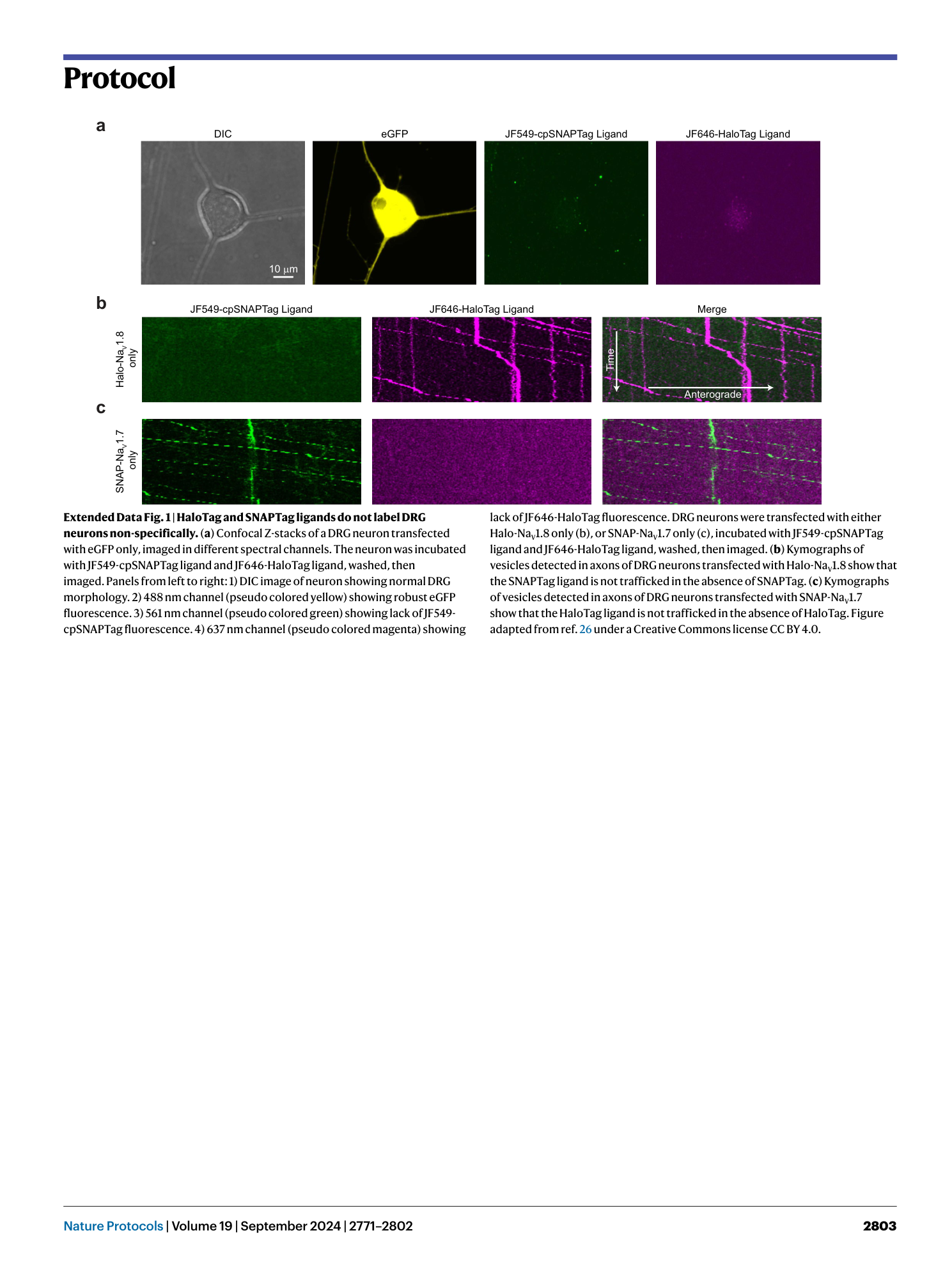
Extended Data Fig. 1 HaloTag and SNAPTag ligands do not label DRG neurons non-specifically.
( a ) Confocal Z-stacks of a DRG neuron transfected with eGFP only, imaged in different spectral channels. The neuron was incubated with JF549-cpSNAPTag ligand and JF646-HaloTag ligand, washed, then imaged. Panels from left to right: 1) DIC image of neuron showing normal DRG morphology. 2) 488 nm channel (pseudo colored yellow) showing robust eGFP fluorescence. 3) 561 nm channel (pseudo colored green) showing lack of JF549-cpSNAPTag fluorescence. 4) 637 nm channel (pseudo colored magenta) showing lack of JF646-HaloTag fluorescence. DRG neurons were transfected with either Halo-Na V 1.8 only (b), or SNAP-Na V 1.7 only (c), incubated with JF549-cpSNAPTag ligand and JF646-HaloTag ligand, washed, then imaged. ( b ) Kymographs of vesicles detected in axons of DRG neurons transfected with Halo-Na V 1.8 show that the SNAPTag ligand is not trafficked in the absence of SNAPTag. ( c ) Kymographs of vesicles detected in axons of DRG neurons transfected with SNAP-Na V 1.7 show that the HaloTag ligand is not trafficked in the absence of HaloTag. Figure adapted from ref. 26 under a Creative Commons license CC BY 4.0.
Supplementary information
Supplementary Information
Supplementary Methods
Reporting Summary
Supplementary Video 1
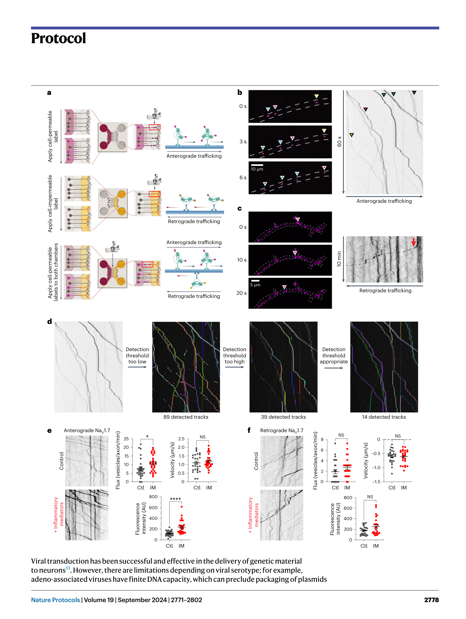
OPAL imaging enables direct visualization of vesicles carrying Halo-tagged membrane proteins with high signal to noise ratio . The use of bright, photostable synthetic fluorophores combined with low background provided an excellent signal-to-noise ratio for clear visualization of vesicles. Anterograde Halo-Na V 1.7 vesicles are visualized using OPAL imaging. This high-resolution imaging technique allows visualization of individual Halo-NaV1.7 vesicles with single-molecule resolution. Reprinted with permission from ref. 25, AAAS.
Supplementary Video 2
OPAL imaging can be extended to investigate co-trafficking of 2 proteins in the same vesicles . Two-color time-lapse OPAL imaging was performed using cell-permeable Halo-tag ligand-JF646 (magenta) and SNAP-tag ligand-JF549 (green). White arrowheads indicate vesicles doubly positive for Halo-Na V 1.8 and SNAP-Na V 1.7 as they move anterogradely along the axon outlined in white. Magenta and green arrowheads indicate vesicles that are singly positive for Halo-Na V 1.8 or SNAP-Na V 1.7, respectively. As described previously, the ∼ 1 μm separation of the SNAP and Halo signals is because of the continued motion of the vesicle during the time between acquisition of images in separate channels. When the vesicle is moving, the SNAP signal is always “behind” the Halo signal because the SNAP (561 nm) channel is acquired first. When the vesicle stops, however, the two signals overlap completely. From ref. 26 under a Creative Commons license CC BY 4.0.
