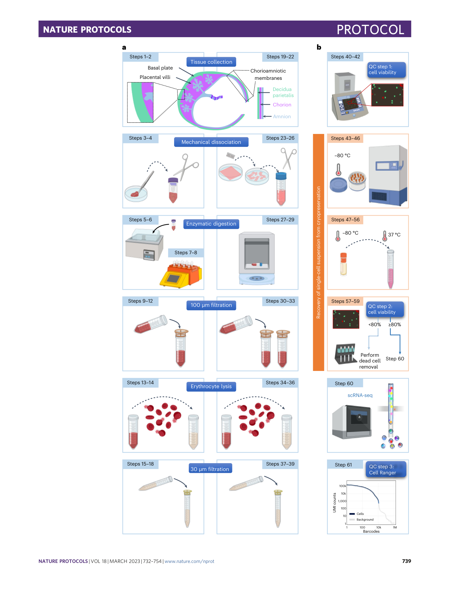
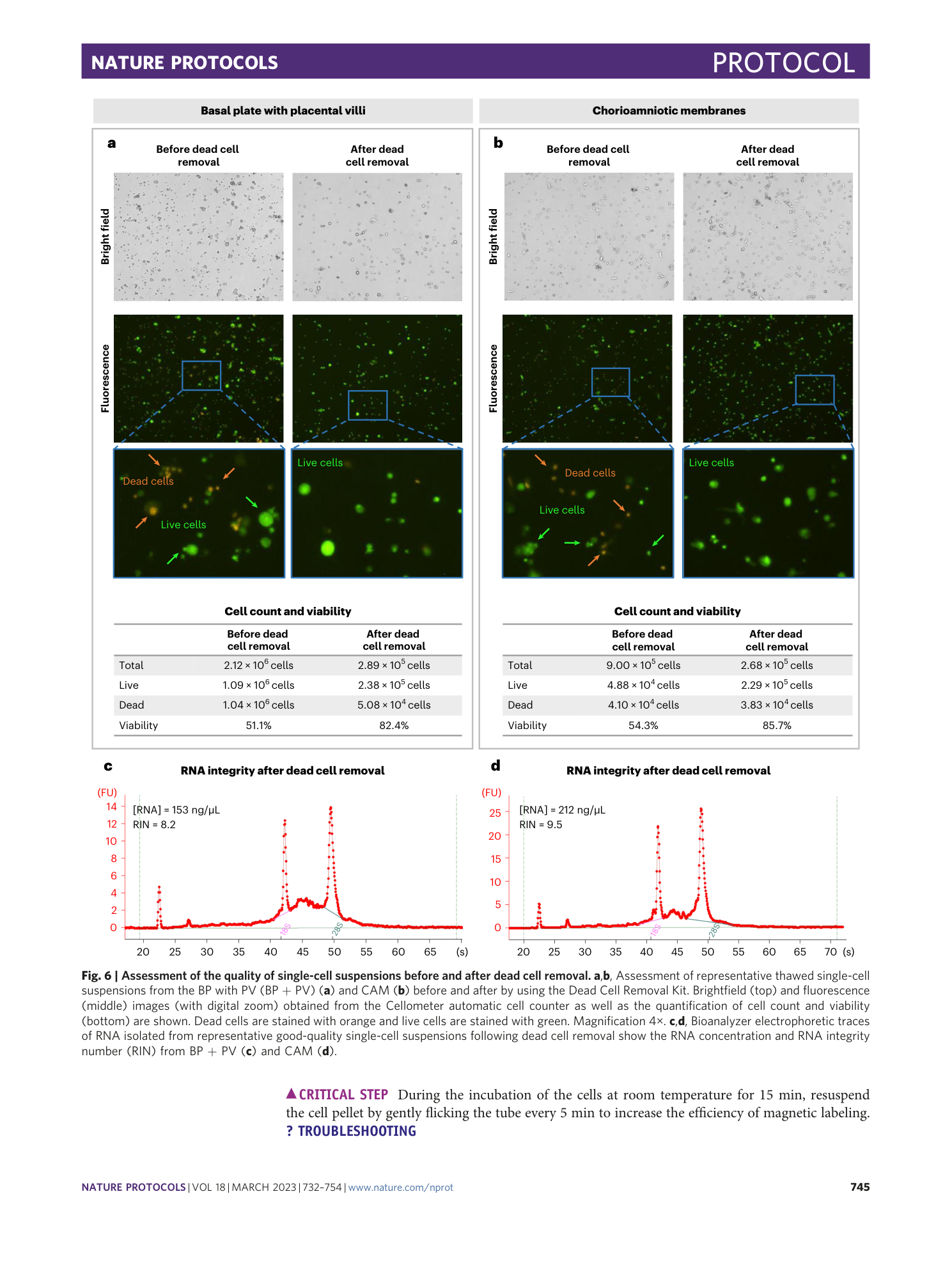
Preparation of single-cell suspensions from the human placenta
Valeria Garcia-Flores, Yi Xu, Errile Pusod, Roberto Romero, Roger Pique-Regi, Nardhy Gomez-Lopez

Extended
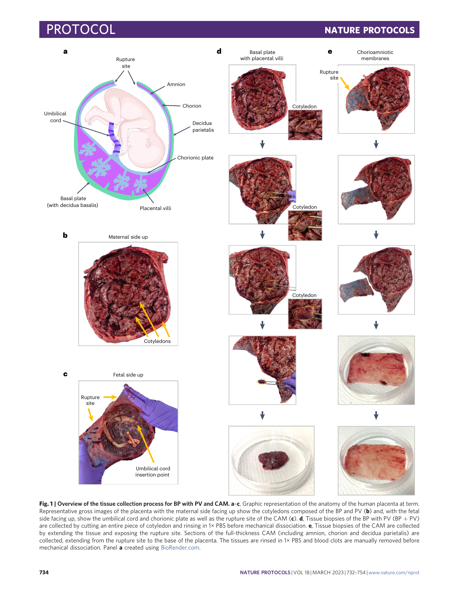
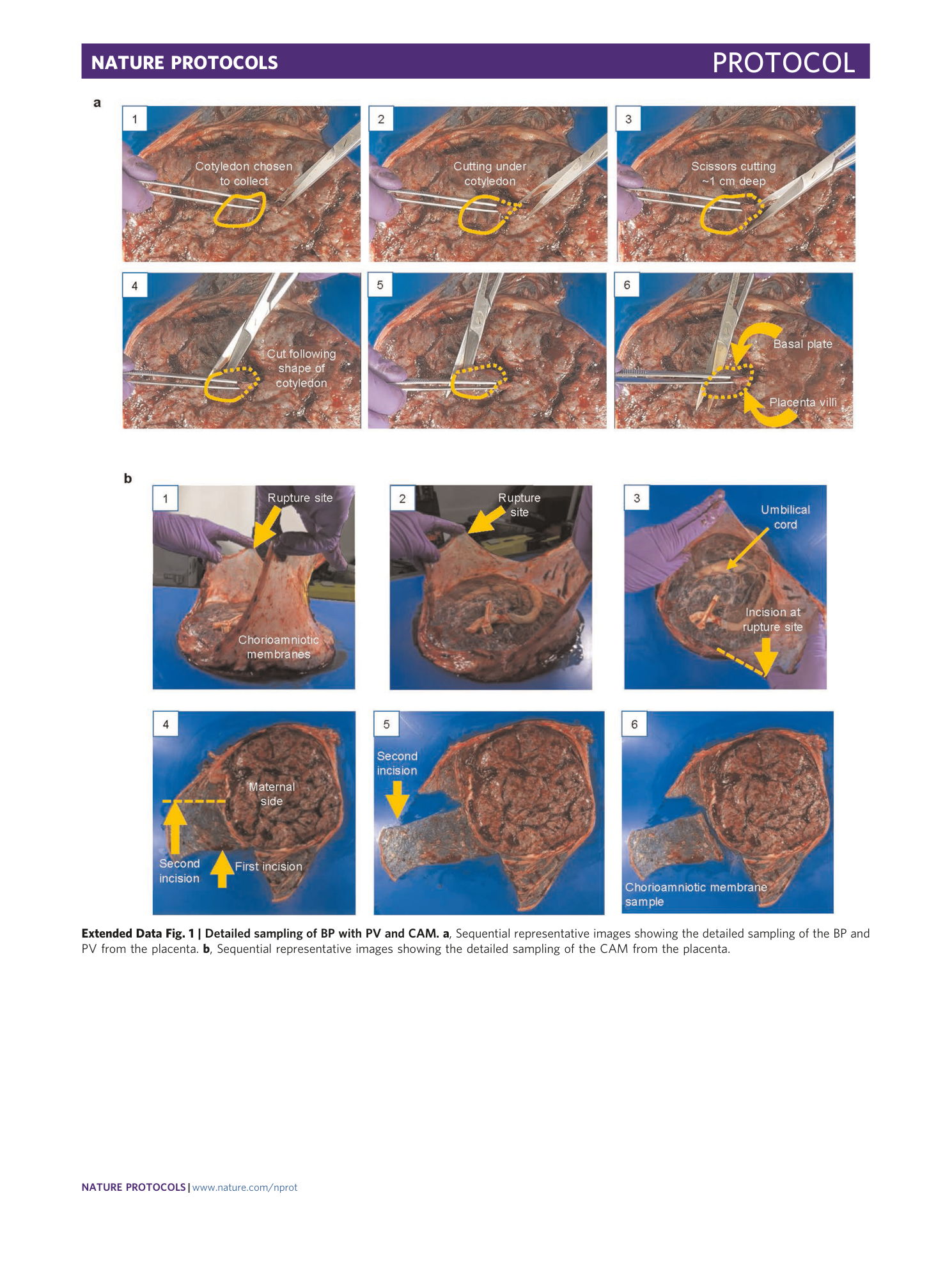
Extended Data Fig. 1 Detailed sampling of BP with PV and CAM.
a , Sequential representative images showing the detailed sampling of the BP and PV from the placenta. b , Sequential representative images showing the detailed sampling of the CAM from the placenta.
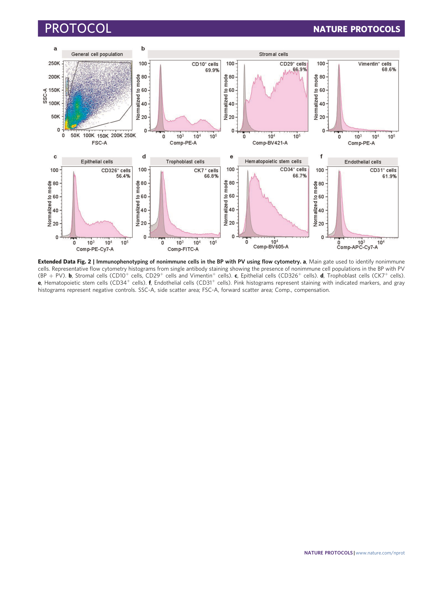
Extended Data Fig. 2 Immunophenotyping of nonimmune cells in the BP with PV using flow cytometry.
a , Main gate used to identify nonimmune cells. Representative flow cytometry histograms from single antibody staining showing the presence of nonimmune cell populations in the BP with PV (BP + PV). b , Stromal cells (CD10 + cells, CD29 + cells and Vimentin + cells). c , Epithelial cells (CD326 + cells). d , Trophoblast cells (CK7 + cells). e , Hematopoietic stem cells (CD34 + cells). f , Endothelial cells (CD31 + cells). Pink histograms represent staining with indicated markers, and gray histograms represent negative controls. SSC-A, side scatter area; FSC-A, forward scatter area; Comp., compensation.
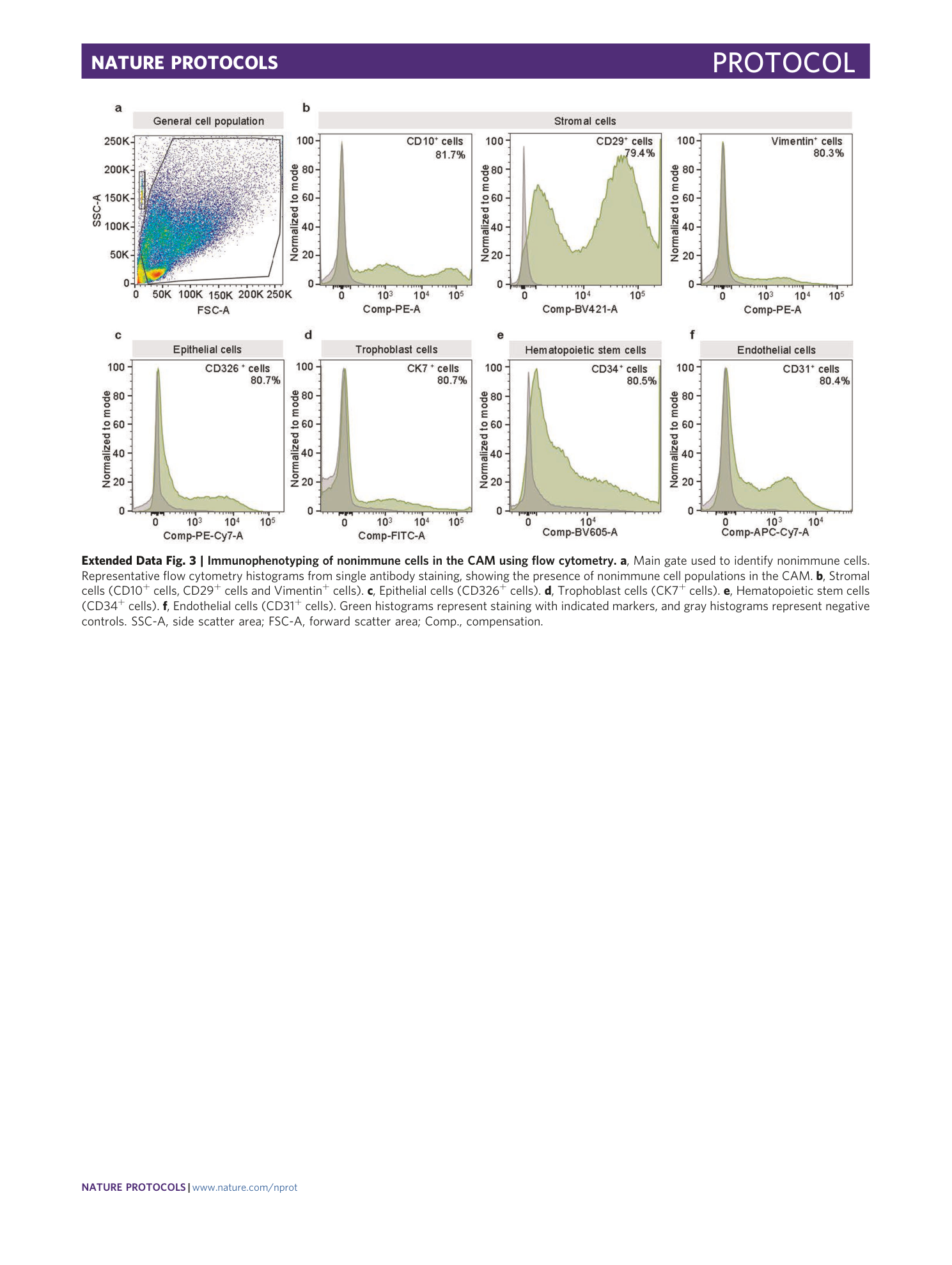
Extended Data Fig. 3 Immunophenotyping of nonimmune cells in the CAM using flow cytometry.
a , Main gate used to identify nonimmune cells. Representative flow cytometry histograms from single antibody staining, showing the presence of nonimmune cell populations in the CAM. b , Stromal cells (CD10 + cells, CD29 + cells and Vimentin + cells). c , Epithelial cells (CD326 + cells). d , Trophoblast cells (CK7 + cells). e , Hematopoietic stem cells (CD34 + cells). f , Endothelial cells (CD31 + cells). Green histograms represent staining with indicated markers, and gray histograms represent negative controls. SSC-A, side scatter area; FSC-A, forward scatter area; Comp., compensation.
Supplementary information
Supplementary Information
Protocol to validate the quality of single-cell suspensions by immunophenotyping.
