Preparation of mouse and human α-synuclein fibrils
Arpine Sokratian, andrew.west west
Abstract
This protocol describes the preparation of homogeneous a-synuclein fibrils. The primary objective is to generate alpha-synuclein fibrils with high homogeneity in size, devoid of other protein species such as monomers and oligomers. The protocol employs shaking to generate the fibrils and sonication to reduce the fibrils to their terminal length. The expected outcome is the production of highly homogeneous fibrils, with a consistent size range of 25±5 nm in radius, and at high concentrations without the presence of monomers and oligomers. This protocol provides a method for producing stable, homogeneous sonicated fibrils that can be stored in batches for in vitro and in vivo experiments. By following this protocol, the fibrils produced will consistently maintain the same size and concentration, thereby ensuring the reliability and reproducibility of subsequent experimental results.
Steps
Generation of α-synuclein fibrils via shaking cycles
Thaw down an aliquot of α-synuclein monomer stock (track down a batch number with identified EU number, concentration, A260/280 ratio) on iceOn ice /quick water bath incubation at 42°C with shaking
Spin down an aliquot of α-synuclein monomer stock solution (20000x g, 0h 10m 0s``4°C ) and measure the concentration using nanodrop
Add 3µL of 10x diluted aliquot in PBS onto nanodrop pedestal;
Parameters:
-
other proteins; coefficient extinction: 5.98; MW: 14.4 kDA (for wild-type human α-synuclein)
-
other proteins; coefficient extinction: 7.45; MW: 14.4 kDA (for wild-type mouse α-synuclein)
Perform two measurements and confirm <10% standard error between two measurements
If necessary, prepare 20X and 30X dilutions to confirm findings.
Equipment
| Value | Label |
|---|---|
| NanoDrop™ One/OneC Microvolume UV-Vis Spectrophotometer | NAME |
| UV-Vis Spectrophotometer | TYPE |
| Thermo Scientific | BRAND |
| ND-ONE-W | SKU |
| https://www.thermofisher.com/us/en/home.html | LINK |
Calculate the reaction mix: 5mg/mL concentration of α-synuclein in 200µL in PBS
Use protein low-binding tubes:
Incubate the reaction at 1000rpmfor 120h 0m 0s using program settings: 1 min ON; 1 min OFF
Equipment
| Value | Label |
|---|---|
| Eppendorf Thermomixer C Model 5382 | NAME |
| Thermomixer C | TYPE |
| Eppendorf | BRAND |
| 5382000023 | SKU |
Equipment
| Value | Label |
|---|---|
| ThermoTop® | NAME |
| Smart block | TYPE |
| Eppendorf | BRAND |
| 5308000003 | SKU |
Spin down the insoluble fraction at 15000rpm,10°C; remove 100µL of supernatant and add 1mL of fresh PBS to the pellet

Gently resuspend and spin down the fibrils at 15000rpm,10°C; remove around 900µL of supernatant (be careful, avoiding disturbing insoluble fraction) and add 1mL of fresh PBS to the pellet (repeat 3 times)
Equipment
| Value | Label |
|---|---|
| Centrifuge 5425/5425 R - Microcentrifuge | NAME |
| Centrifuge | TYPE |
| Eppendorf | BRAND |
| 5406000240 | SKU |
| https://www.eppendorf.com | LINK |
Take out 900 ul of PBS leaving about 100 ul of fibril pellet in the tubes. Transfer the fibrils into 0.2 mL PCR tubes (thick wall). *** spin down the PCR tubes using bench-top centrifuge
Equipment
| Value | Label |
|---|---|
| Mini-centrifuge | NAME |
| Centrifuge | TYPE |
| Fisher | BRAND |
| S67601B | SKU |
| Any standard mini centrifuge with adapters for different tube sizes will suffice | SPECIFICATIONS |
Sonicate the generated fibrils using water bath sonicator at 10C, 30% amplitude and for 1 hour (no OFF ON cycles). Check the level of water in the water-bath is in a line with the tube content.
Sonication
")

Size range and concentration measurements
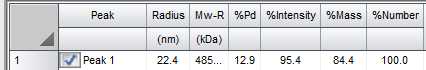
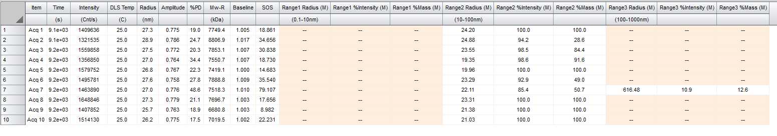
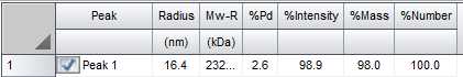
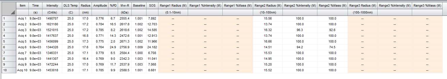
Measure size of sonicated particles using DLS. Indication that α-synuclein fibrils are sonicated to the terminal size can be extrapolated from the distribution of size populations after 10 aqs. 5 sec apart DLS measurement.
-
There is one single population of size in a range of 10-100 nm
-
Average radius is 20-30 nm for human fibrils and 15-25 for mouse fibrils
-
%Mass is no less 95% for the size population from 10-100 nm
See examples below


Measure the concentration of sonicated fibrils:
prepare serial dilutions: 2x; 4x; 8x in 3M Guanidine HCL
Start with preparation of 2x dilution: 4 uL of sonicated fibrils + 4 ul of 6M GuHCL
4x: add 4 uL of 2x diluted sample + 4 uL of 3M GuHCL
8x: add 4 uL of 4x diluted sample + 4 uL of 3M GuHCL
Incubate for 5 min at RT
Add 3 uL of diluted sample onto nanodrop piedestal;
Parameters: other proteins; coefficient extinction: 5.98; MW: 14.4 kDA for human fibrils
Parameters: other proteins; coefficient extinction: 7.45; MW: 14.4 kDA for mouse fibrils
- Confirm that the standard error between the two measurements is less than 10%.
- Calculate the stock concentration by multiplying the measured concentration by the dilution factor
Equipment
| Value | Label |
|---|---|
| NanoDrop™ One/OneC Microvolume UV-Vis Spectrophotometer | NAME |
| UV-Vis Spectrophotometer | TYPE |
| Thermo Scientific | BRAND |
| ND-ONE-W | SKU |
| https://www.thermofisher.com/us/en/home.html | LINK |
