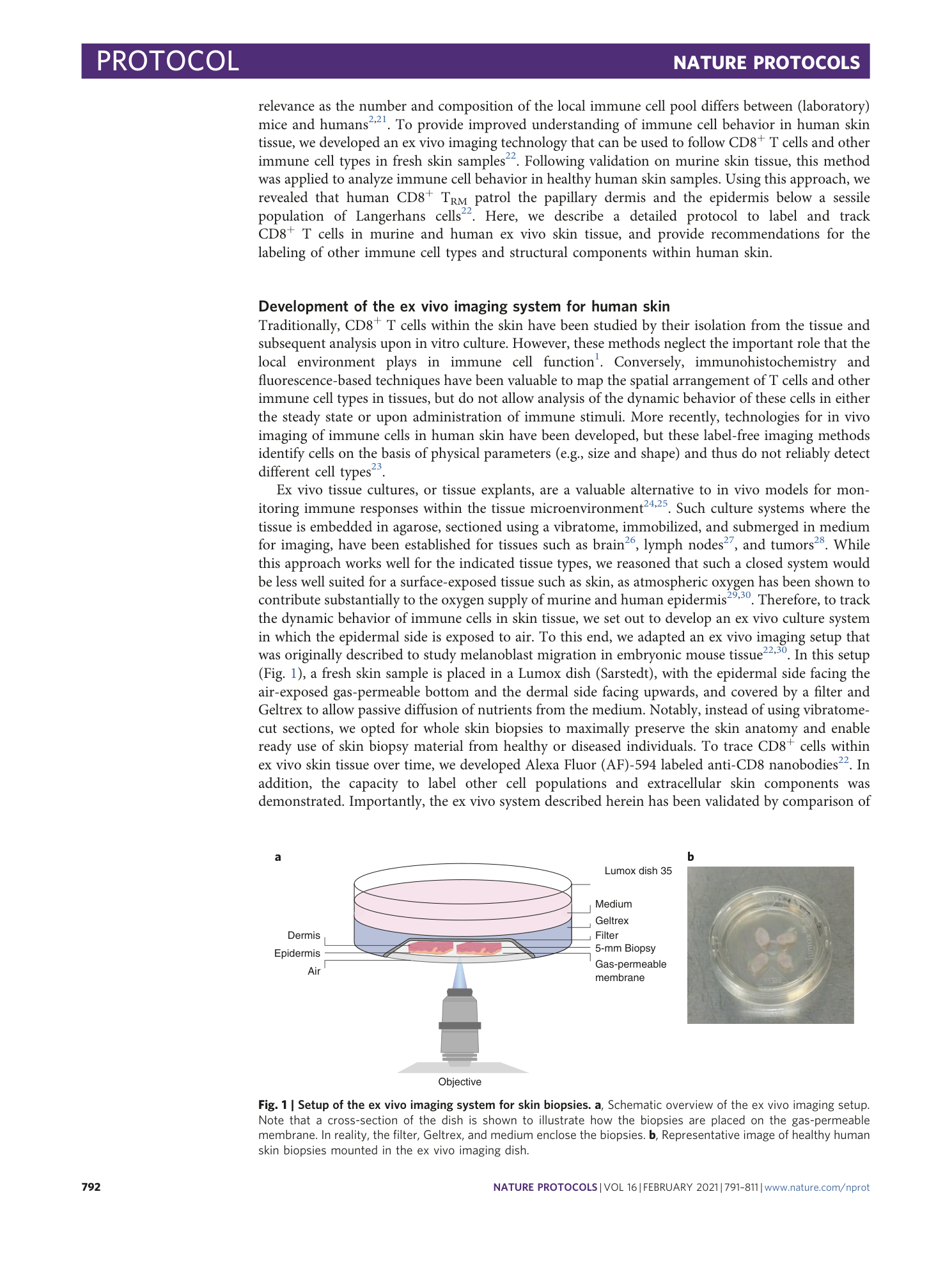
Labeling and tracking of immune cells in ex vivo human skin
Feline E. Dijkgraaf, Mireille Toebes, Mark Hoogenboezem, Marjolijn Mertz, David W. Vredevoogd, Tiago R. Matos, Marcel B. M. Teunissen, Rosalie M. Luiten, Ton N. Schumacher


Supplementary information
Reporting Summary
Supplementary Video 1
CD8 + T cells in murine ex vivo skin regain motility and dendricity after overnight culture. Maximum confocal microscopy projection of OT-I GFP + CD8 + T cells (green) in mouse skin imaged directly after mounting in ex vivo imaging setup. Cells start off round and immobile and regain mobility and dendricity over time (time in minutes). N=2 recordings and 2 independent observations. Scale bar, 50 μm.
Supplementary Video 2
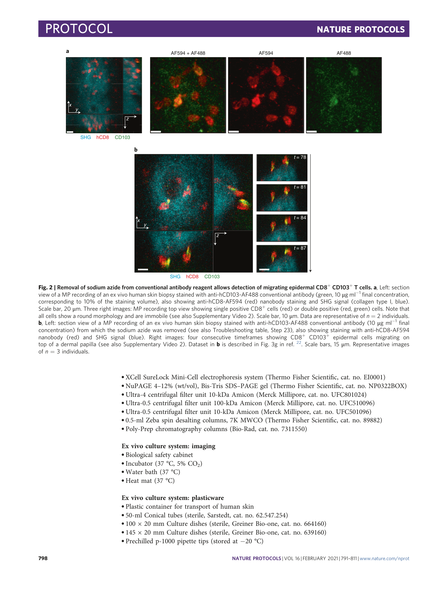
Removal of sodium azide from conventional antibody reagent allows detection of migrating epidermal CD8 + CD103 + T cells in human skin. First segment: Section view of MP recording showing ex vivo human skin biopsy stained with anti-hCD103-AF488 conventional antibody (green) without removal of sodium azide, also showing anti-hCD8-AF594 nanobody staining and second harmonics generation signal (SHG, blue). Note that CD8 + T cells display a round morphology and are immobile. Representative of n=2 individuals. Scale bar, 20 μm. Second segment: Section view of MP recording showing ex vivo human skin biopsy stained with anti-hCD103-AF488 conventional antibody (green) after removal of sodium azide, also showing anti-hCD8-AF594 nanobody staining and second harmonics generation signal (SHG, blue). Note that CD8 + CD103 + T cells display a variable morphology and migrate. Data in second segment adapted from Supplementary Video 11 in 22 . Representative of n=3 individuals. Scale bar, 15 μm.
Supplementary Video 3
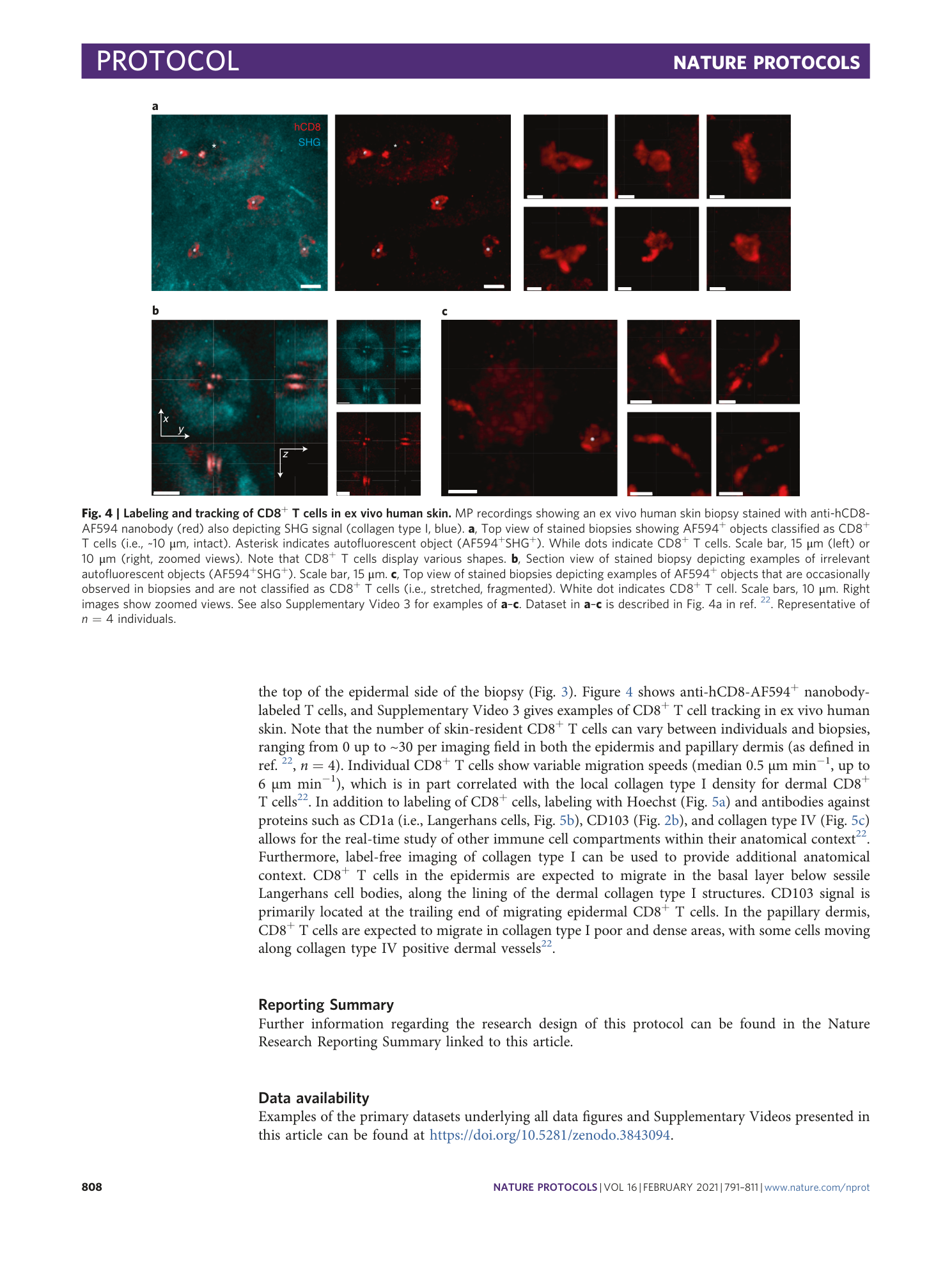
Labeling and tracking of CD8 + T cells in ex vivo human skin. First segment: Top view of MP-recordings showing examples of tracked anti-hCD8-AF594 nanobody labeled T cells (red) that migrate in ex vivo human skin. Second segment: Examples of autofluorescent and fragmented AF594 + objects in ex vivo human skin biopsies stained with anti-hCD8-AF594 nanobody (red), also showing second harmonics generation signal (SHG, collagen type I, blue). Both segments: lines indicate tracked cells. Datasets are described in Fig. 4a in 22 . Data representative of n=4 individuals. Scale bars, 10 μm.
Supplementary Video 4
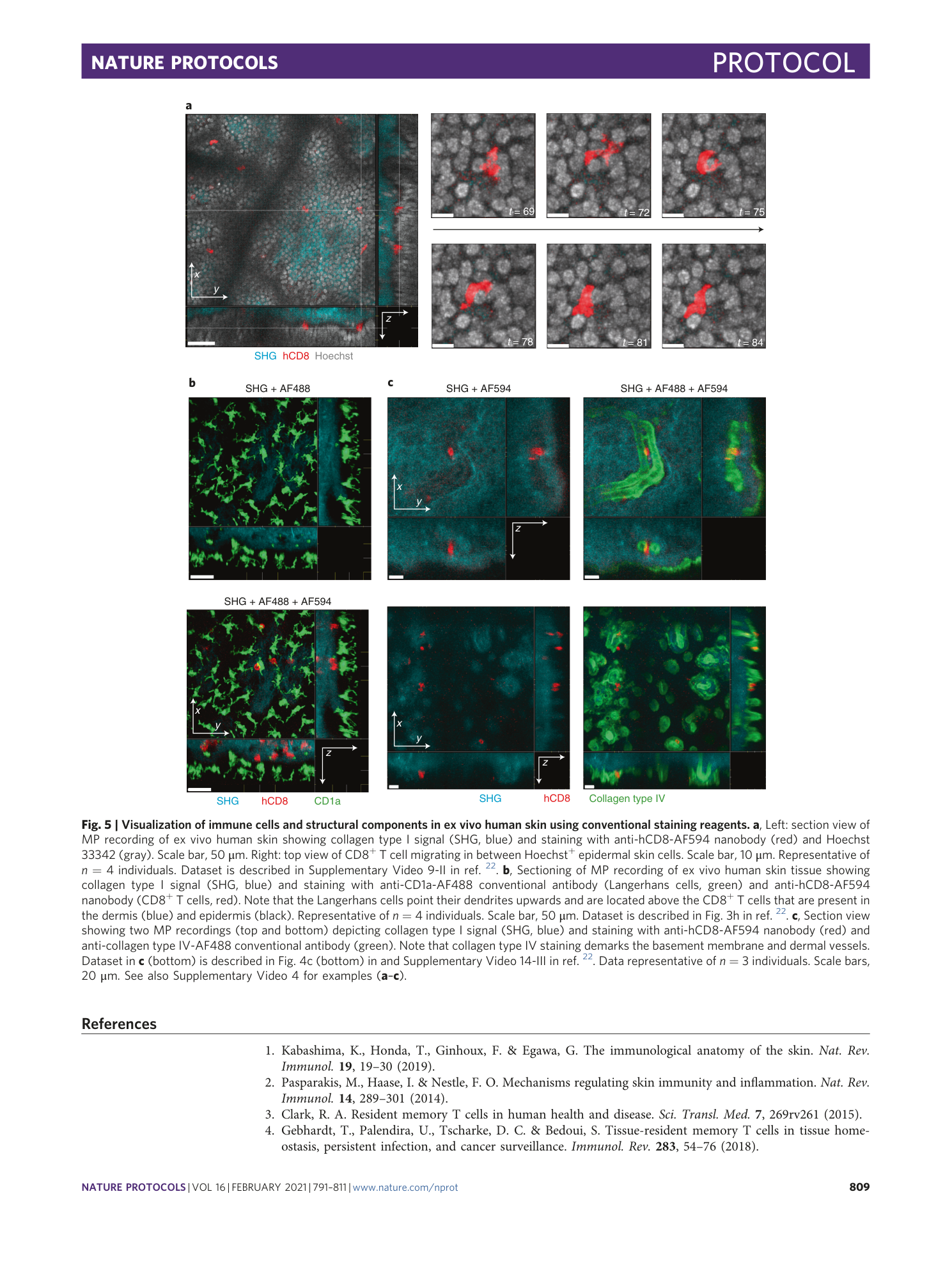
Visualization of immune cells and structural components in ex vivo human skin using conventional staining reagents . First segment: Top view of MP-recording showing anti-hCD8-AF594 nanobody-labeled CD8 + T cell (red) migrating in between Hoechst + nuclei (gray) in the epidermis. Dataset described in Supplementary Video 9-II in 22 . Representative of n=4 individuals. Scale bar, 10 μm. Second segment: Section view showing anti-hCD8-AF594 nanobody-labeled CD8 + T cell (red) migrating below anti-hCD1a-AF488 antibody labeled Langerhans cells (green), also showing second harmonics generation signal (collagen type I, blue). Dataset described in Supplementary Video 12-I in 22 . Representative of n=4 individuals. Scale bar, 50 μm. Third segment: Section view of MP-recording showing anti-hCD8-AF594 nanobody labeled CD8 + T cells (red) migrating alongside anti-human collagen type IV positive structures (i.e., basement membrane and dermal vessels, green), also showing second harmonics generation signal (collagen type I, blue). Dataset is described in Supplementary Video 14-II in 22 . Representative of n=3 individuals. Scale bar, 20 μm.
Supplementary Data 1
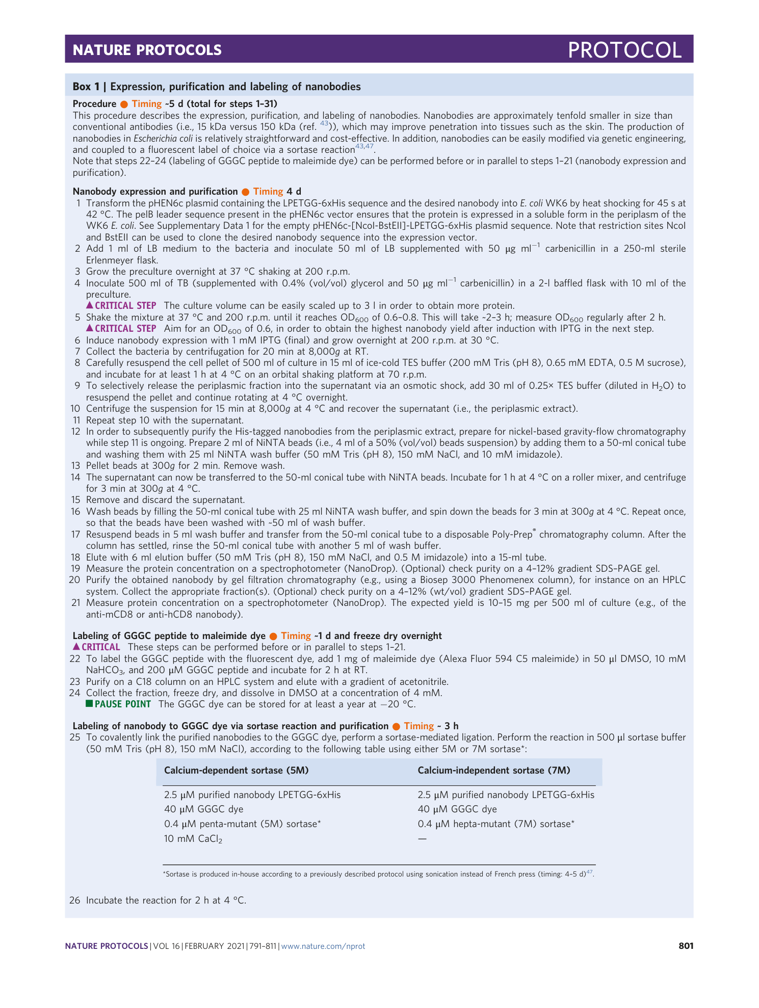
DNA sequence of the empty pHEN6c expression vector containing the LPETGG (i.e., Sortag) and 6xHis (i.e., Histag) sequence and the NcoI and BstEII cloning sites, which can be used to insert the desired nanobody sequence.
