LRRK2 Immunofluorescent staining 

sherbst

Published: 2022-07-25 DOI: 10.17504/protocols.io.n92ldze17v5b/v1

Abstract

Protocol for immunofluorescent staining for LRRK2 in cultured cells using the MJFF2 (c41-2) antibody.

Steps

1.

Culture cells as usual on cover slips or in a plate suitable for imaging.

If using coverslips, we recommend Ø13 mm for cells cultured in 24-well plates.

Note
The MJFF2 (c41-2) antibody only detects concentrated LRRK2. Therefore, controls should be included during the sample preparation to ensure signal specificity. We recommend including a LRRK2 KO or knock down control and a positive control such as 30 min treatment with 1mM LLOMe (H-Leu-Leu-OMe•HBr, 4000725.0001, Bachem) or 50 μM chloroquine (C6628-25G, Sigma-Aldrich).

2.

Fix cells in 4 % PFA/PBS at 4°C for 0h 15m 0s.

3.

Optional: at this step, coverslips or plates can be stored at 4 °C in PBS. Never allow samples to try out.

4.

Permeabilise the plasma membrane by incubating samples in ice-cold MeOH for 0h 10m 0s. This can be done on the bench or the whole plate can be put into the -20 °C freezer. Make sure that samples are always submerged.

5.

Wash samples once in PBS

6.

Incubate samples in blocking buffer for 0h 20m 0s at Room temperature

7.

Incubate samples in primary antibody solution ( MJFF2 (c41-2) antibody diluted 1:100 in blocking buffer) for 1h 0m 0s at Room temperature

Note
At this step, additional primary antibodies can be added to assess LRRK2 localisation. For lysosomal location in mouse samples, we recommend anti-LAMP1 (#1D4B, Developmental Studies Hybridoma Bank) used at 1:100 dilution. Always control for cross-reactivity.

8.

Wash samples three times in PBS

9.

Incubate samples in secondary antibody solution ( eg anti-rabbit-Alexa Fluor™ 488 diluted 1:800 in blocking buffer) for 0h 45m 0s at Room temperature in the dark

Note
If additional primary antibodies were used, also include additional secondary antibodies here (eg anti-rat-Alexa Fluor™ 657).

10.

Wash samples twice in PBS

11.

Incubate sample in DAPI staining solution for 0h 10m 0s at Room temperature in the dark

12.

Wash samples twice in PBS

13.

Mount coverslips onto slides using a mounting medium of choice (eg DAKO Fluorescence Mounting medium) and let try at RT in the dark.* If using well-plates, add PBS to plates.

  • Keep samples in the dark and store them at 4 °C for short-term storage.

    Citation
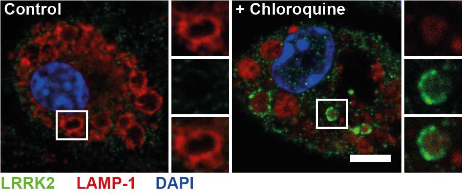
    Murine bone marrow-derived macrophages were treated with 50 μM chloroquine for 30 min and stained for LRRK2 according to the above protocol. LRRK2 accumulates at the lysosomal membrane in response to chloroquine but is not visible in untreated cells using this staining method.
    Murine bone marrow-derived macrophages were treated with 50 μM chloroquine for 30 min and stained for LRRK2 according to the above protocol. LRRK2 accumulates at the lysosomal membrane in response to chloroquine but is not visible in untreated cells using this staining method.

推荐阅读

Nature Protocols
Protocols IO
Current Protocols
扫码咨询