Human Pregnant Uterine Myometrium Tissue Collection and Preservation Methods - UCSD Female Reproductive TMC
Scott Lindsay-Hewett, Valentina Stanley, Louise Laurent, Mana Parast
Abstract
Human pregnant uterine myometrium tissue collection and storage protocol for HuBMAP's UCSD Female Reproductive TMC.
Steps
Preparation
At the time of c-section, obtain a full-thickness uterine myometrial biopsy from the superior edge of the incision site, prior to closure (about 2.0cm x 1.0cmx 0.5cm). Collect the tissue into an empty sterile cup.
Wash the tissue in cold PBS.
Place tissue on the guaze pad keeping the serosal (smooth) side pointing up and endomyometrial side pointing down. Take a photo. Split the tissue in half horizontally, dividing the serosal side from the endomyometrial side.
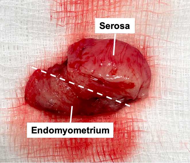
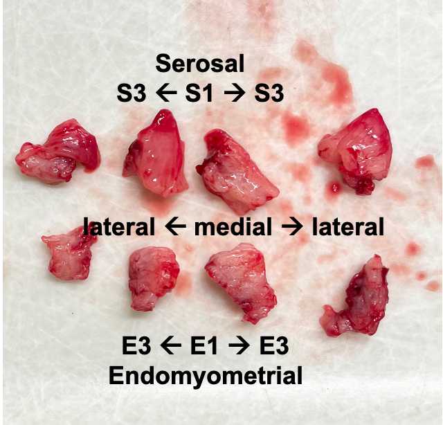
Cut each side into smaller chunks, maintaining the medial to lateral and serosal to endomyometrial orientation.
Preserving the serosal side
Collect cross sections of the serosal side into 10% formalin (x1), RNAlater (x3) and to be snap frozen (x3). The pieces should be about 5mm each, depending on the total size of the tissue. If there is an excess of tissue, collect more of each type. S1 should be most medial, then S2, then S3 should be most lateral.
Preserving the endomyometrial side
Collect cross sections of the endomyometrial side into 10% formalin (x1), RNAlater (x3) and to be snap frozen (x3). The pieces should be about 5mm each, depending on the total size of the tissue. If there is an excess of tissue, collect more of each type. E1 should be most medial, then E2, then E3 should be most lateral.
Flash-freezing
Drop the snap frozen labeled tubes into liquid Nitrogen. Leave for ~2-10 minutes, then store in -80C freezer.
Processing with RNAlater
Place RNAlater tubes into a 4C fridge. Let these sit for 24-48 hours, then remove the RNAlater with a sterile transfer pipette and store in a -80C freezer.
Formalin fixing
After one week in formalin, place each formalin-fixed piece into a labeled cassette to be processed into FFPE blocks.
