High-performance liquid chromatography with electrochemical detection of monoamine neurotransmitters and metabolites
Stephanie J Cragg, Emanuel F Lopes
Abstract
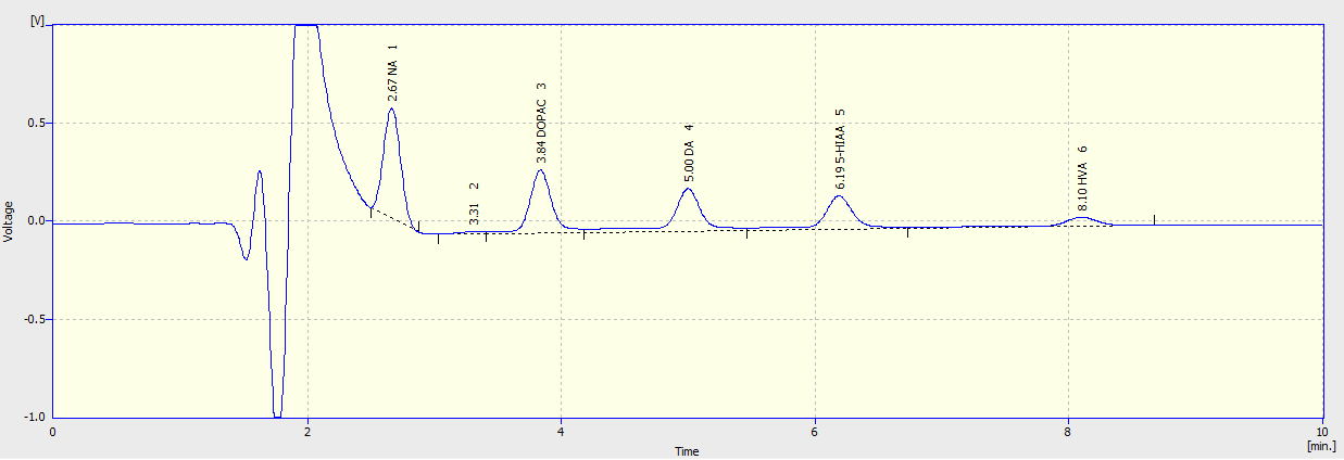
This protocol describes a method to identify and quantify the content of the monoamine neurotransmitters dopamine (DA) and norepinephrine (NE), along with several related metabolites (3,4-Dihydroxyphenylacetic acid – DOPAC; homovanillic acid – HVA; 5-Hydroxyindoleacetic Acid – 5-HIAA) from brain tissue samples using HPLC with electrochemical detection.
Before start
Note 1: This protocol is optimised for detecting tissue punches from acute coronal striatal slices, normally obtained after ex vivo fast-scan cyclic voltammetry experiments (see Protocol: Fast-scan cyclic voltammetry to assess dopamine release in ex vivo mouse brain slices). Tissue obtained from other preparations are suitable with modifications to this protocol, but note the analysis will have to be adjusted.
Note 2 : All reagents, chemicals and solvents must be HPLC grade where possible, or of the highest purity. All solutions must use ultrapure water.
Note 3: To help increase the longevity of the equipment, we keep the HPLC pump flow rate at 0.25mL/min and the electrochemical cell turned off when the HPLC set-up is not in use. When running samples, remember to turn the cell on, and increase the flow rate to 1 mL/min.
Note 4: Do not let air bubbles enter the system. Make sure there is plenty of mobile phase available during sample runs, if the equipment is in between uses the mobile phase can be recycled. When switching to a new batch of mobile phase be sure to turn the pump off when moving the in-flow tubes to prevent air from entering the system.
Steps
Obtaining tissue punches
Dispense 200 µL of 0.1 M PCA solution (see Materials ) into Eppendorf tubes. We generally use each Eppendorf to prepare material from 2 tissue punches.
Lay acute coronal striatal slices (slice thickness is 300 µm) flat on a cutting mat, and use a micro punch to remove a region of the striatum to analyse.
Use forceps to transfer the punch(es) into the Eppendorfs, making sure the punches are submerged in the PCA solution.
Store punches in -80°C until ready to analyse.
Preparing mobile phase
Use a 2 L volumetric flask to make up the Mobile Phase (see Materials ).
Check the pH of the mobile phase is ~4.65; if not, add orthophosphoric acid or sodium hydroxide.
Filter solution with a vacuum pump and PES membrane filter to remove larger contaminating particles.
A day prior to running samples, switch to new mobile phase solution. Ensure no air bubbles enter the system when switching to new mobile phase solution as this can damage the column. Leave overnight to guarantee that the new mobile phase is entirely occupying the system.
When running samples make sure the flow rate is 1 mL/min. Run time should be between 10-20 minutes. Injection volume is 50 µL, check this is the case on the autosampler.
On the DECADE II software, make sure electrochemical cell is on and at 0.7 V. Turn on auto zero.
Preparing and Running Standard Solutions
Make up 10 mM (10-2 M) of each standard solution: 5-HIAA, NA, DOPAC, HVA, and DA (see Materials ).
Proceed to make serial dilutions for each standard solution to reach a final volume of 1 mL of 10-7 M for each standard solution. In addition, prepare a mixed 10-7 M solution containing all of the above compounds.
Run each standard separately, then the mixed standard, followed by a PCA-only sample.
Ascertain retention times for each compound of interest, and confirm that sample components do not co-elute.

Preparing Samples
Defrost samples and homogenize them using handheld sonicating probe.
Centrifuge samples in 4°C at 15,000g for 15 mins, then place back on ice.
If using a pair of 2 mm tissue punches, dilute supernatant by 1:10 in 0.1 M PCA. A pair of 1.2 mm tissue punches can be run undiluted.
Running Samples
Create a new protocol on Clarity software. Make a note of column being used, mobile phase composition, flow-rate, pump pressure, and room temperature.
Aliquot 120 µL of each sample into labelled HPLC vials (with insert).
Load all samples into autosampler. Include 10-7M mixed standard at the beginning, middle, and end of the run to control for degradation.
Data Analysis
Extract data for each sample and the mixed standards.
Calculate metabolite content as pmol/mm3.
Calculate average peak values for each metabolite from each mixed standard.
For each metabolite, based on extracted peak values, calculate pmol of metabolite
present in injected volume (50 µL). Convert this to metabolite present in full volume (200 µL).
If samples were diluted (see above for DLS punches in which the supernatant was diluted 1:10), account for dilution.
If using 2 tissue punches per sample, divide by 2.
This results in a value in pmol/punch, convert to pmol/mm3.

