GeoMX Digital Spatial Profiler Whole Transcriptome Assay for Human Pancreas
Jing Chen, Martha Campbell-Thompson, Clayton E Mathews, Ann Fu, Yanping Zhang, Heather Kates, Alberto Riva
pancreas
FFPE
GeoMX
Nanostring
whole transcriptome assay
morphology
insulin
cytokeratin
CD31
fluorescence
hybridization
nCounter
HuBMAP
TMC-PNNL/UF
RNAsequencing
transcriptomics
DNA
UF ICBR
bioinformatics
Illumina
spatial transcriptomics
islet
exocrine pancreas
endothelium
duct
Disclaimer
Refer to Nanostring online user manuals for additional information (https://nanostring.com/support/nanostring-university/).
Abstract
Purpose:
This protocol details processing fixed paraffin sections from human pancreas for the Nanostring GeoMX digital spatial profiler (DSP) whole transcriptome assay (WTA). This technology is based upon hybridization of samples with RNA reagents composed of >20,000 probes for full coverage of protein covering mRNA. The RNA probes are designed with complimentary RNA sequences and UV cleavable linkers and DSP barcodes. Morphology markers are used to identify cell types of interest with nuclear counterstaining permitting tissue spatial information. Sample regions of interest (ROI) are defined based on the 4 morphology markers and each ROI is illuminated with UV light thereby cleaving hybridized RNA probes. Cleaved probes are aspirated via microcapillary and deposited into a unique well of a 96-well collection plate. The collection plate is used for next generation sequencing (NGS).
Scope:
This document was written following GeoMX- NGS guidelines (university.nanostring.com) with minor modifications for the University of Florida Molecular Pathology Core.
The entire workflow is described for manual slide staining and GeoMX DSP instrument use. Steps for library construction, sequencing, bioinformatics, and data analysis are in outline form only. Library preparation and sequencing are outsourced to the University of Florida ICBR facility.
Expected outcome:
RNA suitable for bulk RNA-sequencing will be obtained from fixed human pancreas sections within regions of interest and subareas defined by cell morphology markers.
Before start
Preheat waterbath to 37°C.
Prepare 1x Antigen Retrieval Solution and preheat in steamer while slides are dewaxing.
Steps
This procedure details processing of paraffin slides, in set of 4, for GeoMX WTA by manual methods. Methods using the Leica BOND autostainer are detailed on the Nanostring website.
Paraffin blocks containing 4% paraformaldehyde fixed human pancreas are screened by H&E staining and histopathology review https://www.protocols.io/view/human-pancreas-histopathology-assessment-n92ld956xg5b/v1.
Wash steps in this protocol may be abbreviated such as one wash= 1 x and three washes= 3 x.
Room temperature (RT) is used except where different temperature is noted.
Slide preparation
Cut paraffin block to fresh sample.
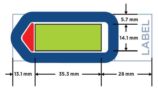
Section at 5 µm and place sections centrally on Superfrost Plus slides as shown in Fig. 1. Tissue samples should have maximum width and length of ~14 and 36 mm, respectively, for inclusion within the GeoMX instrument slide holder gasket. Tissue outside the gasket region should be carefully removed without creating folds or leaks to gasket seal.
Air dry mounted slides overnight before user or storage.
Recommended storage is for < 2 weeks in a desiccator at 4°C.

Bake slides at 60°C for 1h 0m 0s immediately before use.
Slide deparaffinization
Dewax and rehydrate sections using slide tray stations or a Leica XL slide autostainer.
Do not allow tissue sections to dry once rehydrated and avoid touching tissue when drying around edges.
Xylene 3 x 0h 5m 0s. CitriSolv or another xylene substitutes can be used.
100% Ethanol 2 x 0h 5m 0s
95% Ethanol 2 x 0h 5m 0s
PBS 1 x 0h 1m 0s
Antigen retrieval
Prepare Antigen Retrieval Solution (1x Tris EDTA, pH 9) in Coplin jar.
Pre-heat in steamer.
Transfer slides to Antigen Retrieval Solution jar and steam for 0h 20m 0s.
Immediately transfer slides to PBS.
Wash slides in PBS 1x 0h 5m 0s.
Slides can be stored in PBS up to 1h 0m 0s.
RNA retrieval
RNA retrieval solutions needed:
Proteinase K working solution: Place in Coplin jar and prewarm to 37°C for 0h 15m 0s in water bath.
NBF stop solution in two Coplin jars
PBS wash
Incubate slides in Proteinase K jar at 37°C for 0h 15m 0s in water bath.
Wash PBS 1 x 0h 5m 0s. Proceed to next step immediately.
Post-retrieval fixation for pancreas
Incubate in 10% NBF for 0h 5m 0s.
Incubate in NBF stop buffer 2 x 0h 5m 0s.
Wash PBS 1 x 0h 5m 0s.
Slides can be stored in PBS wash solution up to 1h 0m 0s at RT or up to 6 hrs at 4°C.
RNA probe hybridization and stringency washes
Prepare RNA probe mixture in dedicated RNAse-free area away from nCounter work or other NGS workflows.
Warm buffer R to RT before opening vial.
Repeat steps 8.5-7 for each slide.
Close slide tray, insert into hybridization oven, and clamp in place.
Incubate for 16h 0m 0s at 37°C. Slides must remain under humid conditions to prevent drying out of hybridization solution. The chamber can be placed in a ziplock bag for additional security.
Thaw RNA Probe Mix on ice. Before use, mix thoroughly by pipetting. Store unused RNA detection probes at 4°C up to 6 months or re-freeze.
Make RNA probe hybridization solution as per Table 1. Before use, briefly vortex and quick spin down.
| A | B | C | D |
|---|---|---|---|
| Buffer R (ul) | RNA Probe Mix (ul) | DEPC Water (ul) | Final Volume (ul) |
| 200ul x N | 25ul x N | 25ul x N | 250ul x n |
| 800 | 150 | 50 | 1000 |
Table 1. RNA probe hybridization solution preparation for NGS readout. Amounts per slide are shown. Volumes needed for 4 slides are provided.
Prepare slide hybridization incubator with RNAse AWAY and dry or rinse with DEC water. Place Kimwipes on bottom and wet with 2 x SCC or DEPC water to create humid environment during overnight hybridization.
The hybridization chamber can be a key point of contamination by oligo probes.
Remove each slide from PBS, tilt and wipe off excess PBS, then place in slide holder of hybridization chamber.
Work quickly to avoid tissue drying.
Immediately add 200 ul RNA probe solution to each slide. Do not introduce air bubbles during pipetting.
Apply a Hybrislip over each section by gently laying down over tissue, starting from one edge, and avoiding introduction of air bubbles.
Post-hybridization stringency washes.
Solutions needed:
2xSSC
2xSSC-T
2x SSC/50% formamide
Warm 100% formamide to RT before opening.
Prepare 2x SSC/50% formamide by mixing 100ml each of 4x SSC and 100% formamide.
Add solution to 2 Coplin jars and preheat to 37°C in water bath .
Dip slides in 2x SSC to remove coverslips.
If they do not come off immediately, move to 2x SSC-T for maximum of 0h 5m 0s.
If coverslip have not come off in 2x SSC-T, move slides to stringent washes where coverslips should come off.
Do not force off coverslips to avoid tissue damage.
Wash in 2xSSC/50% formamide 2 x 0h 25m 0s at 37°C in water bath.
Wash in 2xSSC 2 x 0h 5m 0s.
After last wash, slides can be stored in 2xSSC for up to 1h 0m 0s.
Carefully wash all containers coming into contact with the hybridization solution (Proteinase K, formamide mixture 2XSSC) with RNAse AWAY to prevent probe contamination. Use separate Coplin jars for different probe mixtures.
Morphology marker incubation
Incubate sections with up to 3 morphology markers (MM) and one DNA counterstain to define cell types of interest. Morphology markers are validated by in-house testing using the RNA assay conditions.
Clean humidity chamber with RNAse AWAY.
Thaw nuclear marker, vortex then spin down for at least 1 minute to pellet any precipitates. Pipette from top of vial only. Close tightly after use and refreeze at -20°C.
Remove one slide from 2xSSC and tap clean on stacked clean towels to remove excess buffer.
Cover section with 200ul Buffer W for 0h 30m 0s in dark slide chamber while preparing morphology marker solution. Make sure Buffer W extends 2-3 mm past section borders.
Prepare MM solution. Mix solution by flicking followed by quick spin down.
| A | B | C | D | E | F | G |
|---|---|---|---|---|---|---|
| Nuclear Marker | MM1 | MM2 | MM3* | Buffer W** | Total Volume | |
| Slide n | 22 ul x n | 5.5 ul x n | 5.5 ul x n | 5.5 ul x n | 187ul x n | 220 ul x n |
| 1 | 22 | 5.5 | 5.5 | 0 | 187 | 220 |
| 2 | 44 | 11 | 11 | 374 | 440 | |
| 3 | 66 | 16.5 | 16.5 | 561 | 660 | |
| 4 | 88 | 22 | 22 | 748 | 880 |
Table 2. Morphology marker solution preparation. Volumes are in ul for up to 4 slides. *Optional third MM. Adjust Buffer W amount when using 3 MM. MM: morphology marker.
Working with one slide at a time, remove Buffer W by tapping slide edge on stacked towels then add 200ul morphology marker solution/slide in humidity chamber. Repeat for each slide.
Incubate slides in dark slide chamber for 1h 0m 0s
Tap slide edge on towels to remove MM solution then wash slides in 2x SSC 2 x 0h 5m 0s.
Submerge slides in 2x SSC for 2 x 0h 5m 0s. Load slides in DSP within 6h 0m 0s of last wash for optimal results.
Alternatively, store slides in 2xSSC for up to 2 days at 4°C in dark.
Slides must be stored in dark to avoid cleavage of DSP tags by UV light.
GeoMX DSP run
Start run by selecting New/Continue Run under Data Collection in the GeoMx DSP Control Center.
Clean the bottom of slide with 70% ethanol and place slide in slide holder with slide label towards front of instrument.
Complete loading slides then lower slide tray clamp. Cover sections with 6 ml Buffer S. Record slots for each slide.
Load a 96-well aspiration plate in DSP. Check solution containers are filled and waste container is empty.
Follow prompts in the GeoMx DSP Control Center to identify slides.
Name slides according to protocol https://dx.doi.org/10.17504/protocols.io.n2bvj6dnblk5/v1
Include UF HuBMAP sampleID and morphology marker abbreviations in order of channels followed by date (MMDDYY). Example: P1-21_DNA_PanCK_INS_CD31_121021.
Fill in the GeoMX Seq Code plate to be used with the plate barcode in the Readout Group Information grid.
Click update button and finalize and download readout package buttons to USB drive.
Upload unzipped configuration files to DSP kits used for Morphology and WTA kits used in slide preparations (nanostring/dsconfigfiles). Config files must match names of assays used on slides.
Define scan area for each sample using x- and y-sliders and scan. The minimum tissue slide filters particles and small tissue areas from scan. The sensitivity slider adjusts intensity of tissue identification.
Draw regions of interest (ROI) using geometric, segmented or other tools.
Maximum RO1 size is ~ 650 µm diameter circle.
Segment ROIs for AOI (area of illumination) for cell types of interest based on positive or negative selection by morphology markers. Minimum of ~100 nuclei/AOI is recommended for sufficient RNA.
Insulin is used to designate beta-cells in the endocrine compartment, CD31 is used to define endothelium, and panCK is used to define ductal epithelium in the exocrine compartment. Acinar cells are defined by the lack of staining for CD31 and PanCK in the exocrine ROI.
Exit scan workspace and approve ROI to start ROI collection. 2h 0m 0s
After ROI/AOI collections are completed, follow prompts to remove slide holder and collection plate.
Remove buffer from each slide with pipette and dispose. Open clamps and unload slides. Store slides in 2x SSC at 4°C up to 7 days.
Clean slide holder as directed and replace in instrument. Follow prompts to close instrument door.
Slide reuse
RNA assay slides can be restained following UV exposure for 3 minutes to strip tags from bound probes followed by washes using with 2xSSC-T. Proceed with additional staining.
NGS library and sequencing
Transfer collection plate to the University of Florida ICBR Gene Expression and Genotyping Core or other NGS core facility for library preparation and Illumina sequencing.
Refer to Nanostring user manual for additional information.
Library will be prepared after indexing PCR with pooling and purification.
Library will be evaluated for quality and fragment size distribution using a BioAnalyzer 2100 (Agilent).
Core facility will perform Illumina sequencing.
Core facility will process Illumina FASTQ sequencing files and transfer back to laboratory.
Data analysis
Share FASTQ files and annotation file with data analyst who will analyze RNAseq data using R scripts for GeoMx DSP workflow (Bioconductor - GeoMxWorkflows). Data analysis may also be performed with GeoMX DSP software on auxiliary server.
Manage annotations (tags). Add columns to further define regions and cell types from morphology markers.
Run various testing including QC, data scaling, normalization, background, statistical tests, and ratios.
Analyze in spatial context using spatial deconvolution software and scRNAseq data.
Complete analysis and upload data to HuBMAP HIVE.

