Fontana-Masson staining
Csilla Novák, Andrés Mauricio Jaramillo Flautero, Matthias Prigge, Cristian González-Cabrera, Celine Winter
Abstract
The Fontana-Masson Staining Protocol is a detailed method for staining free-floating sections (20-50 microns) to detect melanin and other pigments in tissue samples. The procedure involves preparing a silver nitrate working solution, treating sections with triton, and sequential incubation in pre-heated silver nitrate solution, gold chloride, and thiosulfate solutions, with thorough washing steps between each incubation. The process concludes with sections being placed in PBS for mounting and counterstaining. This protocol is critical for histological studies in biology, particularly for pigment analysis in tissues.
Steps
Fontana-Masson staining
Use free floating sections between 20-50 microns.
Silver Nitrate working solution:
-
Prepare a 1:3 solution of silver nitrate solution in distilled water. Prepare enough solution for the amount of sections you want to stain.
-
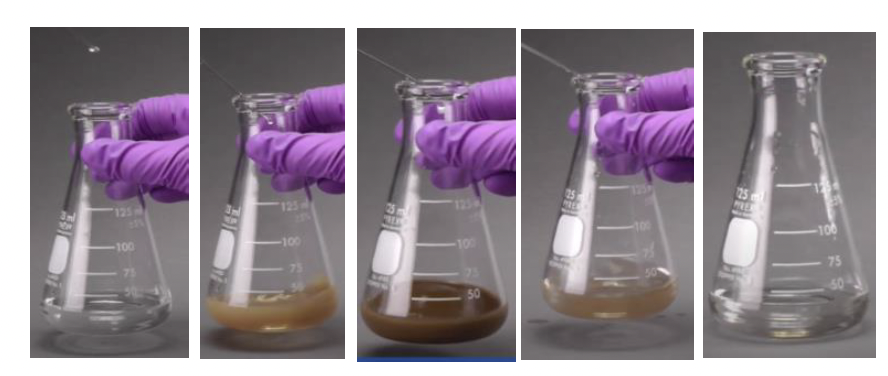
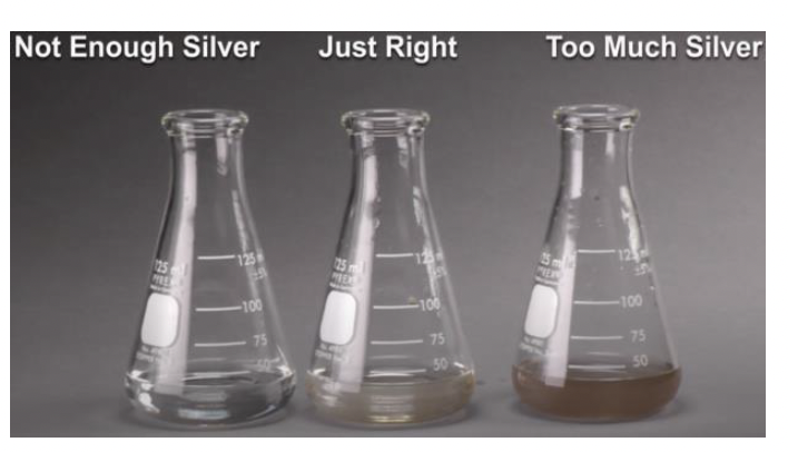
Add drop by drop concentrated ammonium hydroxide, while shaking, until the solution gradually turns transparent. Be careful and stop the addition of ammoniacal solution as soon as you get transparency.
Place the sections to stain (up to 7-8 sections) in a 2mL Eppendorf tube. Incubate the sections in 0.3% triton for two hours at room temperature while shaking
Wash 3 times with distilled water for 5 min each.
Pre-heat the silver nitrate working solution for 5 minutes at 60°C.
Incubate the sections in the preheated solution for 20 minutes at 60°C.
Wash the sections 3 times with abundant distilled water.
From now on, perform the following steps in individual sections:
Incubate the section with gold chloride solution. Apply enough solution to cover the section (approximately 100-150µL). Manually shake the tube for 30 seconds.
Wash the section 3 times with abundant distilled water.
Incubate the section with thiosulfate solution. Apply enough solution to cover the section (approximately 100-150µL). Manually shake the tube for 2 minutes.
Wash with tap water for 2 minutes.
Wash 3 times with distilled water for 5 min each.
Place the sections in PBS for mounting, counterstaining, etc.
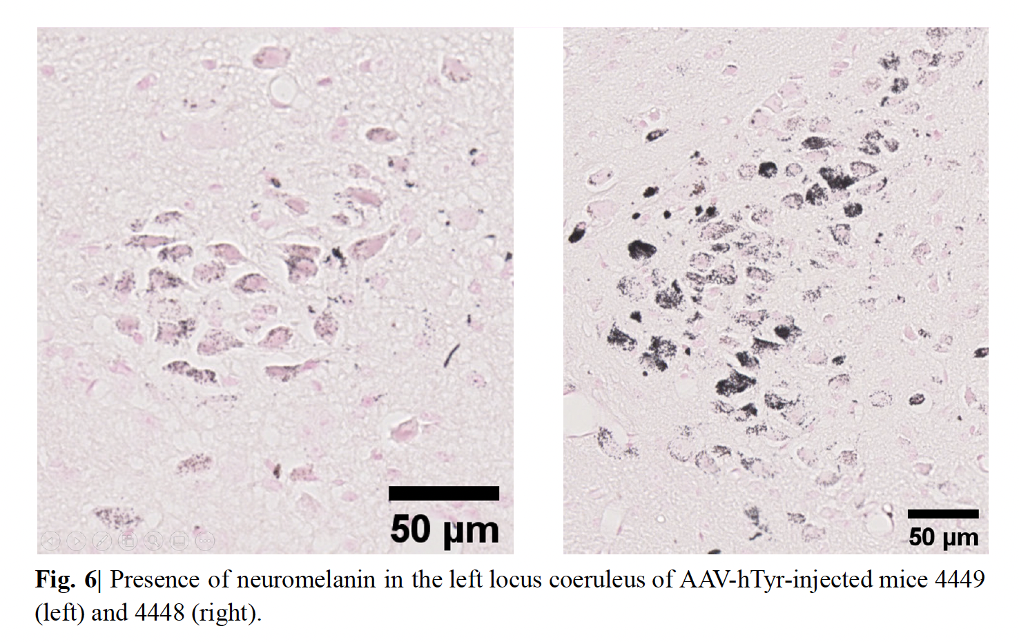
Results
Slices should now show an amplified pigmentation as show in figures