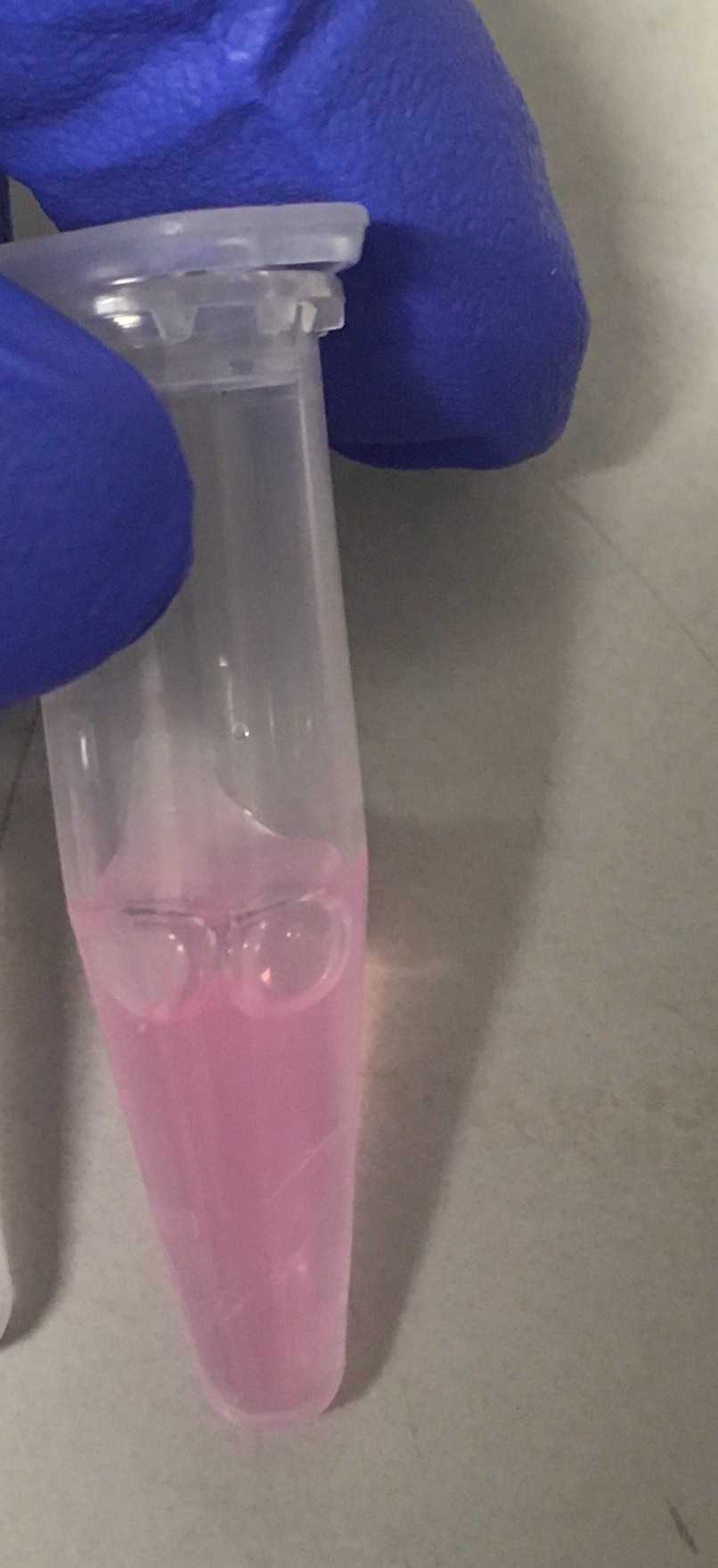
FPCount protocol - in-lysate (purification free) protocol
Eszter Csibra, Guy-Bart Stan
Abstract
FPCount is a complete protocol for fluorescent protein calibration, consisting of:
-
FP expression and production of cell lysates.
-
FP concentration determination in a microplate reader.
-
FP fluorescence quantification in a microplate reader.
Results can be analysed with the corresponding R package, FPCountR.
This in-lysate version of the protocol uses the ECmax protein quantification protocol of FPs in lysates and does not require His-tag purification of the FPs . Note that it is only suitable for FPs with entries in FPbase. If you want to verify or validate results, it's recommended you follow the 'short' protocol, which requires FP purification, or the 'complete' protocol, which requires FP purification and compares three protein quantification methods.
Summary
-
Expression
-
Harvesting/Washing
-
Lysis
-
Fractionation
-
Protein concentration and buffer exchange
-
Quantification of FP concentration (part1)
-
Quantification of FP fluorescence
-
Protein storage
-
Calibration of Plate Reader
Before start
This in-lysate version of the protocol uses the ECmax protein quantification protocol of FPs in lysates and does not require His-tag purification of the FPs . Note that it is only suitable for FPs with entries in FPbase. If you want to verify or validate results, it's recommended you follow the 'short' protocol, which requires FP purification, or the 'complete' protocol, which requires FP purification and compares three protein quantification methods.
Steps
Expression
[ Day 1 ]
Overnight culture set-up:
- 50ml LB
- 50ul cam
- 50ul arabinose 0.02%
- glycerol stock scraping of BL21/pS381_ara_His-FP transformant (or equivalent)
- 30oC 250rpm
- overnight expression…
Harvesting and Washing
[ Day 2 ]
Buffers:
- Wash buffer = T50N150
- 50 mM Tris-Cl pH 7.5, 150 mM NaCl
- Doesn’t need protease inhibitors
- Can be substituted by T50N300
- Resuspension buffer = T50N300+pi
- 50 mM Tris-Cl pH 7.5, 300 mM NaCl
- protease inhibitors (pi; 1 tablet/10ml)
- filter sterilise as pi makes things cloudy/doesn’t go into solution well
- Lysis Buffer = T50N300+pi
- lysozyme 1X (100ug/ml)
- DNase I (1000U/ml)
- CaCl2 stock
- MgCl2 stock
- Dilution Buffer = T5N300+pi
- For protein assays
- How much buffer will I need?
- T50N150 - maybe 35ml/FP
- T50N300 + pi (f/s) - make master stock > 10ml per FP.
- T50N300 + pi + lysozyme = <3ml per FP
- T50N300 + pi for protein assays = <7ml per FP
- total pi tablets required = < 10ml per FP = 1 pi per FP
Procedure:
- Prechill 1x 50ml falcon tube per FP on ice, 15min
- Prechill 1-2x 50ml falcon tubes per FP on ice - for sonication (choose 1x aliquot if using 20 OD cells)
- Prechill big centrifuge, 4oC
- Remove culture from incubator; for some FPs it will be clear by eye if expression levels are good.
- Transfer to falcon on ice; cool for 20min
- From now on cultures and protein should be kept on ice and spun at 4oC unless otherwise stated.
- Take OD (use 100ul of culture 1:10 in LB)
- Expect maybe 4-6 OD/ml.
- Calculate volume or fraction of total required for 20 OD worth of cells. (20 OD = 1x 20OD/2ml aliquot for sonication)
- Expect fraction to be 0.1-0.2 meaning we’re only using 10-20% of the culture for this calibration. So expecting vol to be 5-10ml.
Example OD calculation:
| A | B | C | D | E | F | G |
|---|---|---|---|---|---|---|
| 40/total OD | 40/total * 50 | |||||
| 1 | mCherry | 0.418 | 4.18 | 209 | 0.19 | 9.57 |
| 2 | eg1 | 0.3 | 3 | 150 | 0.267 | 13.3 |
| 3 | eg2 | 0.6 | 6 | 300 | 0.133 | 6.67 |
- Add 20 OD to the prechilled tubes set aside for aliquotted cultures.
- (Original cultures can stay on ice or be stored in fridge.)
- Spin 3,220xg, 10min, 4oC
- Resuspend in 5ml WASH buffer w pipetboy/5ml stripette; Add 30ml more WASH buffer or so
- Spin 3,220xg, 10min, 4oC
- Resuspend in 2ml (for 20 OD) Lysis Buffer.
- Lysis Buffer = T50N300 + pi + lysozyme. (No DTT for His tag purifs.)
- eg. for 5 FPs, Take 12 ml of T50N300+pi and add 24ul lysozyme.
Lysis
Prep for next stage: pre-chill the microfuge to 4oC.
Lysis by Sonication
- Stand falcon in small plastic beaker full of ice.
- Sonicate: 50% amplitude, 10s on/off, 2min .
- NB. 2min means 2min of sonication. as we’re doing 10s on/off, this takes 4min.
- Solution should go from turbid to clear.
- If you have many FPs, after 6 falcons you have enough sample to fill the microfuge for the next stage - it’s worth starting the DNase step (30min) then the spin (30min) before the other samples are sonicated.
DNase I treatment
Optional step: essential if using A280 assay but not essential for the ECmax assay. Note that DNase I = 31 kDa meaning similar in size to FPs in a way that would affect estimates in Gel1 (though not Gel2 as it shouldn’t bind the column), and is sensitive to vortexing.
- Prepare DNase I stock: 1000 U/ml DNase I in ddH2O
- To lysates in T50N300, add:
- DNase I to 50 U/ml final (20X dilution)
- CaCl2 to 5mM final (13mM ideal for DNase I, <5mM recommended with His resins)
- MgCl2 to 50mM final
- Mix thoroughly
- Reaction: 30min at 4oC
Fractionation
Prep for next stage: prep eppies and cool them for after the spin
[ Day 2 ]
Spin out insoluble fraction.
- Split 2ml lysates into 4x 0.5ml prechilled eppies.
- Spin in prechilled microfuge - 30min 16Kg 4oC.
- Result: 4x 0.5ml of soluble lysate (if using 20 OD)
- Transfer SOLUBLE fractions to new tubes - can mix so that you have fewer eppies.

Prep for next step: Change temp on microfuge to 21oC, and open lid to let it warm up. Proteins should be protected by the protease inhibitors.
Protein concentration (not always optional)
Notes before starting:
- Amicons are not compatible with >0.1% Triton X100. If sonication was used to lyse cells, no problem. If Triton was used to lyse cells, typically this means there’s 0.1% in the lysis buffer, which may affect this step.
- Amicon concentration can be taken as an opportunity for buffer exchange, eg. dilute out protease inhibitors and buffer exchange from T50N300 to T5N15. But obviously to compare different proteins they should be treated the same. Note: for the experiments in the paper, for the in-lysate experiments I did not buffer exchange at all, I merely concentrated lysates in lysis buffer.
Steps:
- Plan:
-
- concentrate lysates 16x in lysis buffer
-
- resuspend to 100ul
- Use Amicon Ultra 10K columns
- 500ul capacity (500ul -> 15ul possible)
- spin at 14Kg 10’ to concentrate
- spin at 1Kg 1’ to recover
- Concentration.
- Add lysate (400ul) to 10K amicon column (1/n)
- 10’ 14Kg spin at 21oC
- expect it to go down to 50ul
- discard flowthrough
- Repeat as many times as needed. (eg. n = 4 works well for me.)
- Recover result (needs to be <50ul)
- turn column over into fresh eppy
- 1’ 1000g
- Take sample, measure volume precisely, dilute back to 100ul+. 100ul is enough for one quantification, 200ul+ will allow for repeats.
FP Calibration in Plate Readers Protocol
The idea behind this protocol is to make most efficient use of protein. Therefore, ideally, one 100ul aliquot of FP is all that is needed for all the fluorescence and concentration assays, and these can be done consecutively on a single dilution series. Currently an ‘exhaustive’ workflow includes 1 fluorescence assay and 3 protein assays but two of these assays can only be done on purified proteins. As this is the ‘in-lysate’ protocol, this only uses 1 fluorescence assay and 1 protein assay - the ECmax assay.
Summary:
- You will need 100ul elution of each FP to be calibrated.
- Access to all plate readers to be calibrated for a clear few hours is ideal, but the time required depends entirely on ambition: the number of instruments, channels and FPs.
- Prep dilutions as 200ul in the same type of plate as you use for bacterial assays -> absorbance scans and fluorescence quants.
Quantification of FP Concentration (part1)
There is only one protein concentration assay ('protein assay') in this simplified workflow - the ECmax assay.
Therefore plates to scan in order are:
- 200ul clear - ECmax assay/fluorescence assay
–
[ Day 2 contd ]
Prepare dilutions of FPs. Run scans in plate reader for FP quantification.
1. Make Protein Dilutions in Eppies
In order to validate the protein quantitation assays, I dilute each FP using serial dilutions and measure the concentration and fluorescence of each. In order to maximise both the dilution series and the number of FPs quantifiable per plate, I typically arrange FPs in 96-well plates in pairs of rows (AB, CD, EF, GH) - each FP is therefore measured as a duplicate. This leaves the columns for the dilution series itself, with 11 used for protein dilutions, and one for the buffer. (NB: In theory, arranging FPs in columns, with 7 dilutions plus one buffer for each FP, would allow for 6 proteins to be measured at the same time.)
I recommend preparing dilution series in 1.5ml eppies: they are easier to handle (and see into) than wells of a deep well plate, important for avoiding errors.
Typical FP dilution series:
| A | B | C | D |
|---|---|---|---|
| 1 | 100 of elution | 900 | 500 |
| 2 | 500 of prev dilution | 500 | " |
| 3 | " | " | " |
| 4 | " | " | " |
| 5 | " | " | " |
| 6 | " | " | " |
| 7 | " | " | " |
| 8 | " | " | " |
| 9 | " | " | " |
| 10 | " | " | " |
| 11 | " | " | " |
2. Fill standard clear 96-well plate
Typical arrangements:
Clear Plate: 200ul per well. 200ul per well.
| A | B | C | D | E | F | G | H | I | J | K | L | M | N | O |
|---|---|---|---|---|---|---|---|---|---|---|---|---|---|---|
| FP1 | row1 | A | dilution1 (neat) | dilution2 (1:2) | d3 | d4 | d5 | d6 | d7 | d8 | d9 | d10 | d11 | buffer |
| row2 | B | dilution1 (neat) | dilution2 (1:2) | d3 | d4 | d5 | d6 | d7 | d8 | d9 | d10 | d11 | buffer | |
| FP2 | C | |||||||||||||
| D | ||||||||||||||
| FP3 | E | |||||||||||||
| F | ||||||||||||||
| FP4 | G | |||||||||||||
| H |
Steps:
- Distribute 200ul buffer into column 12 with multipipette (Dispensing mode; 5ml; 200ul * 8; Asp 4, Disp 5).
- Add protein dilutions to plate with single channel manual pipette. Pipette tip use can be minimised if dispensing from the lowest concentration protein (dilution11) first.
3. Measure absorbance in clear plate - ECmax assay
Prewarm plate reader to temp used for cell growth curve assays to make sure the calibrations are valid for those temperatures - eg. 30oC.
Plate reader runs with clear plate:
| A | B | C | D | E | F |
|---|---|---|---|---|---|
| 1 | instrument1 | clear | none | A200-1000 | 2 min |
Only one scan is needed here. That is an absorbance scan between 200nm and 1000nm. On the Spark this is the maximum range you can do, and you don't need more anyway. The ECmax wavelength will vary, and the high wavelength is required for path length quantification.
Quantification of FP Fluorescence
The purpose of this step is to quantify the fluorescence of each FP dilution in the same plate that the E. coli growth curves will be carried out in .
[ Day 2 contd ]
Measure fluorescence in clear plate
Plate reader runs with clear plate (continued):
| A | B | C | D | E | F |
|---|---|---|---|---|---|
| 1 | instrument1 | clear | seal | FP-dependent fluorescence channel1 | 10min |
| 2 | instrument1 | clear | seal | FP-dependent fluorescence channel2 | 10min |
| 3 | instrument2 | clear | seal | FP-dependent fluorescence channel1 | 10min |
| 4 | instrument2 | clear | seal | FP-dependent fluorescence channel2 | 10min |
Fluorescence data should be gathered on a range of gains (I use 40-120), for one (or more) channels, as required. For eg. while mScarlet is best measured at emission channel 595/35, it can be useful to measure it in 620/20 as well, in cases where the 595/35 channel would pick up too much crosstalk from another FP (such as YFP).
How I name channels (filter sets):
Channels are named as a combination of the excitation, then emission filter, which are given names to indicate the region of the visible spectrum they cover, followed by numbers. eg. the standard GFP filter set is ‘green1green2’, which excites at wavelength 485nm (bandwidth 20nm) and measures emission at wavelength 535nm (bandwidth 25nm).
Protein storage
Proteins should be stored in the fridge protected from the light. Long term storage and re-testing post freeze-thaw has not been tested.
Analysis - Calibration of Plate Reader
The aim of this protocol is that for a given FP, we can relate the number of FP molecules to the 'relative' fluorescence units observed in a given instrument, with a given filter set, and gain. The previous steps described how to purify FPs to produce calibrants and how to run the assays. The output data from such assays must now be analysed to obtain calibrations.
To assist with this process, we have developed an R package called FPCountR. The full details are on the GitHub page. In brief, functions in this package are provided for each analytical step. parser() functions parse plate reader data into idealised formats. get_concentration() functions calculate protein concentration from absorbance data. The generate_cfs() function obtains conversion factors using concentration and fluorescence data. Finally, process_plate() and calc_percell() functions extract data from microbial growth curves in units of molecules, and molecules/cell.

