Extraction and selection of high-molecular-weight DNA for long-read sequencing from Chlamydomonas reinhardtii
Frédéric Chaux-Jukic, Nicolas Agier, Stephan Eberhard, Zhou Xu
Disclaimer
The authors declare no conflict of interest.
Spotlight Video
The video below is a supplement with extra context and tips, as part of the protocols.io Spotlight series, featuring conversations with protocol authors.
#尊敬的用户,由于网络监管政策的限制,部分内容暂时无法在本网站直接浏览。我们已经为您准备了相关原始数据和链接,感谢您的理解与支持。
<iframe title="YouTube video player" src="https://www.youtube.com/embed/JcFiDv2eMpk?si=UXu9R4YO6AwLJywu" height="315" width="560"></iframe>
Abstract
Recent advances in long-read sequencing technologies have enabled the complete assembly of eukaryotic genomes from telomere to telomere by allowing repeated regions to be fully sequenced and assembled, thus filling the gaps left by previous short-read sequencing methods. Furthermore, long-read sequencing can also help characterizing structural variants, with applications in the fields of genome evolution or cancer genomics. For many organisms, the main bottleneck is to develop robust methods to obtain high-molecular-weight (HMW) DNA for whole genome sequencing purposes. We developed an optimized protocol to extract DNA suitable for long-read sequencing from the unicellular green alga Chlamydomonas reinhardtii , based on CTAB/phenol extraction followed by a size selection step for long DNA molecules. We provide validation results for the extraction protocol, as well as statistics obtained with Oxford Nanopore Technologies sequencing.
Before start
On the day of extraction:
-
Set water bath to 65°C (large enough for one 50-ml tube per sample)
-
Chill isopropanol to -20°C (around 2.5 ml per sample)
Steps
Harvesting and storage of cells
Grow Chlamydomonas reinhardtii cells in 100mL TAP medium under low light (~5 µmol photon.m-2.s-1) with constant shaking at 100rpm, or in other conditions as appropriate for the specific experiment.
Harvest the cells at the end of exponential growth phase (~107 cells.mL-1) in 50 mL tubes by centrifugation 4000x g. Discard supernatant.
Store cell pellets at -20°C (optional).
DNA extraction
Thaw cell pellets at Room temperature (not applicable if the pellets were not frozen) and put on ice.
Centrifuge at 4000x g,4°C and discard any liquid left.
Resuspend by gentle pipetting in 3mL of CTAB solution preheated at 65°C.
Add 5µL of proteinase K (stock solution: 20 mg.mL-1) and 5µL of RNase A (stock solution: 100 mg.mL-1), mix by pipetting and incubate at 65°C for 0h 20m 0s in a water bath.
Centrifuge at 4000x g and distribute 3 x 1 mL of each supernatant into 2 mL tubes. Discard pellet.
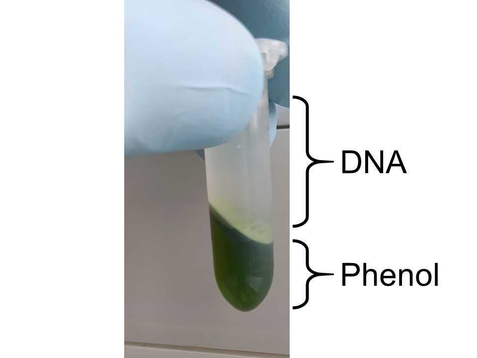
Add 1mL of phenol:chloroform:isoamyl alcool (25:24:1) to each tube, mix gently by inverting 10 times and centrifuge at 20000x g,4°C. Transfer the aqueous (upper) phase to new 2 mL tubes.

Add 1µL of proteinase K and 1µL of RNase A, mix gently by inverting 3 times and incubate at 50°C for 0h 20m 0s.
Add 1mL of chloroform:isoamyl alcohol (24:1) to each tube, mix gently by inverting 3 times and centrifuge at 20000x g,4°C. Transfer the upper phase to new 2 mL tubes.
Add 700µL of isopropanol to each tube and mix gently by inverting 10-12 times. A visible DNA precipitates should form.
Centrifuge at 20000x g,4°C, discard the supernatant carefully, add 500µL of ice-cold 70% ethanol without disturbing the pellet and centrifuge at 20000x g,4°C.
Discard the ethanol carefully by pipetting and eliminate the remaining traces using a SpeedVac for 0h 5m 0s at Room temperature or leave tubes open for 0h 30m 0s at Room temperature .
Resuspend the pellet in 30µL of ultrapure water overnight at Room temperature.
Purification and selection of high-molecular-weight DNA
Pool the samples corresponding to the same initial culture in one 1.5ml Lo-Bind tube, add an equal volume (e.g. 3 x 30µL) of AMPure XP beads, mix gently by hand for 0h 5m 0s.
Place the tubes on a magnetic rack and let the beads aggregate on the side of the tube for 0h 5m 0s. Discard the supernatant. Remove the tubes from the magnetic rack, resuspend the beads in 60µL of ultrapure water for at least 0h 15m 0s at Room temperature.
Place the tubes on a magnetic rack and let the beads aggregate on the side of the tube for 0h 5m 0s. Transfer the supernatant to new 1.5 mL Lo-Bind tubes and discard the beads.
Measure DNA concentration and purity using a Nanodrop and/or a Qubit device.
Adjust each sample to ~150 ng.µL-1 in 60µL of ultra-pure water, gently mix with an equal volume of Short Read Eliminator (SRE) buffer (SRE kit, Circulomics) and centrifuge at 10000x g,4°C. Pipet out the supernatant very carefully as the pellet is colorless and can easily be lost.
Gently add 200µL of freshly prepared 70% ethanol to the pellet. Wash by centrifuging at 10000x g,4°C and discarding the ethanol.
Repeat Step 21.
Air dry the pellet for 0h 10m 0s at Room temperature and resuspend in 50µL of pre-warmed EB buffer (SRE kit, Circulomics) by incubating at 50°C for 0h 20m 0s.
Proceed to long-read sequencing or store at 4°C for later use (at least several weeks). Do not freeze to avoid DNA shearing.
Quality check
DNA purity can be assessed by Qubit or Nanodrop.
Absence of small fragments can be assessed by electrophoresis on agarose gel and size distribution can be assessed by pulsed-field gel electrophoresis (PFGE).

