Chlamydomonas reinhardtii nuclear transformation by electroporation.
João Vitor Molino
Disclaimer
DISCLAIMER – FOR INFORMATIONAL PURPOSES ONLY; USE AT YOUR OWN RISK
The protocol content here is for informational purposes only and does not constitute legal, medical, clinical, or safety advice, or otherwise; content added to protocols.io is not peer reviewed and may not have undergone a formal approval of any kind. Information presented in this protocol should not substitute for independent professional judgment, advice, diagnosis, or treatment. Any action you take or refrain from taking using or relying upon the information presented here is strictly at your own risk. You agree that neither the Company nor any of the authors, contributors, administrators, or anyone else associated with protocols.io, can be held responsible for your use of the information contained in or linked to this protocol or any of our Sites/Apps and Services.
Abstract
This protocols describe the steps required for nuclear transformation of Chlamydomonas reinhardtii by electroporation.
Here you can find a video following the protocol.
Before start
Prepare a ice bucket * Separate cuvettes, keep them on ICE
- Allow linearized vectors to melt
- Keep transformation buffer on ICE/Fridge
- Prepare 50 mL centrifugal tubes with 10 mL TAP medium for recover stage
Steps
DNA Preparation
Digest a large enough amount of plasmid. The goal is to have a concentrated digested sample in the range of 250-700 ng/uL.
- Select the appropiate enzymes for linearization. Usually, restrictions sites in flanking position to the expression cassete.
- Mix all components for digestion
40µg. Digest for6h 0m 0sat37°C. - Column purify digestion (Avoid gel purify, since vector backbone may helps to prevent intracelular DNAses action). * Use a PCR purification kit to purify the digestion reaction.
- Quantitate by absorbance measurement (i.e. Nanodrop).
| A | B |
|---|---|
| Component | Amount |
| 10X Cutsmart NEB | 6.0 uL |
| XbaI | NEB 20 U/uL |
| KpnI HF | NEB 20 U/uL |
| Plasmid 1219.9 ng/uL | 40 uL |
| ddH20, Molecular grade | 8.0 uL |
Typical reaction setup
Cells preparation
Aseptically inoculate 250mL with wild type cells. Either by scrappeing cells of a plate with a inocculating loop or from a previous cultured cells.1. Incubate at 25°C , under constant shaking (~150-180 RPM) and light (60-80 µmols de photons/m2s) until a cell density from 3x 10^6 cells/mL to 6x 10^6 cells/mL is reached.
Pellet cells in centrifuge tubes. Separate culture in sufficient amount of sterile 50mL centrifuge tubes or larger volume tubes, and centrifuge for 2000x g,25°C.
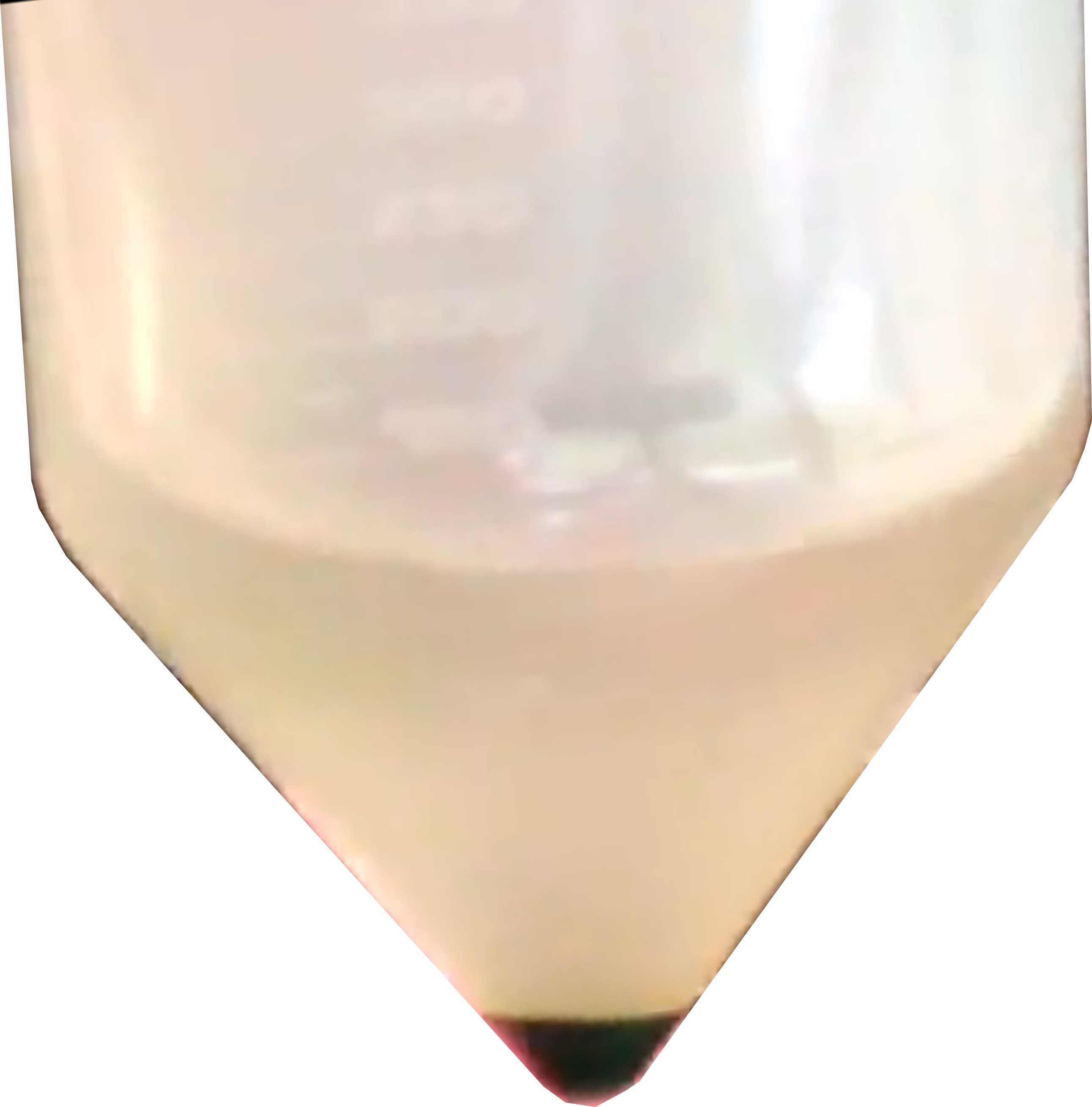
- Genttly ressupend cells at 3-6-108 cells/mL 8 cells/mL in Transformation Buffer.
Transformation
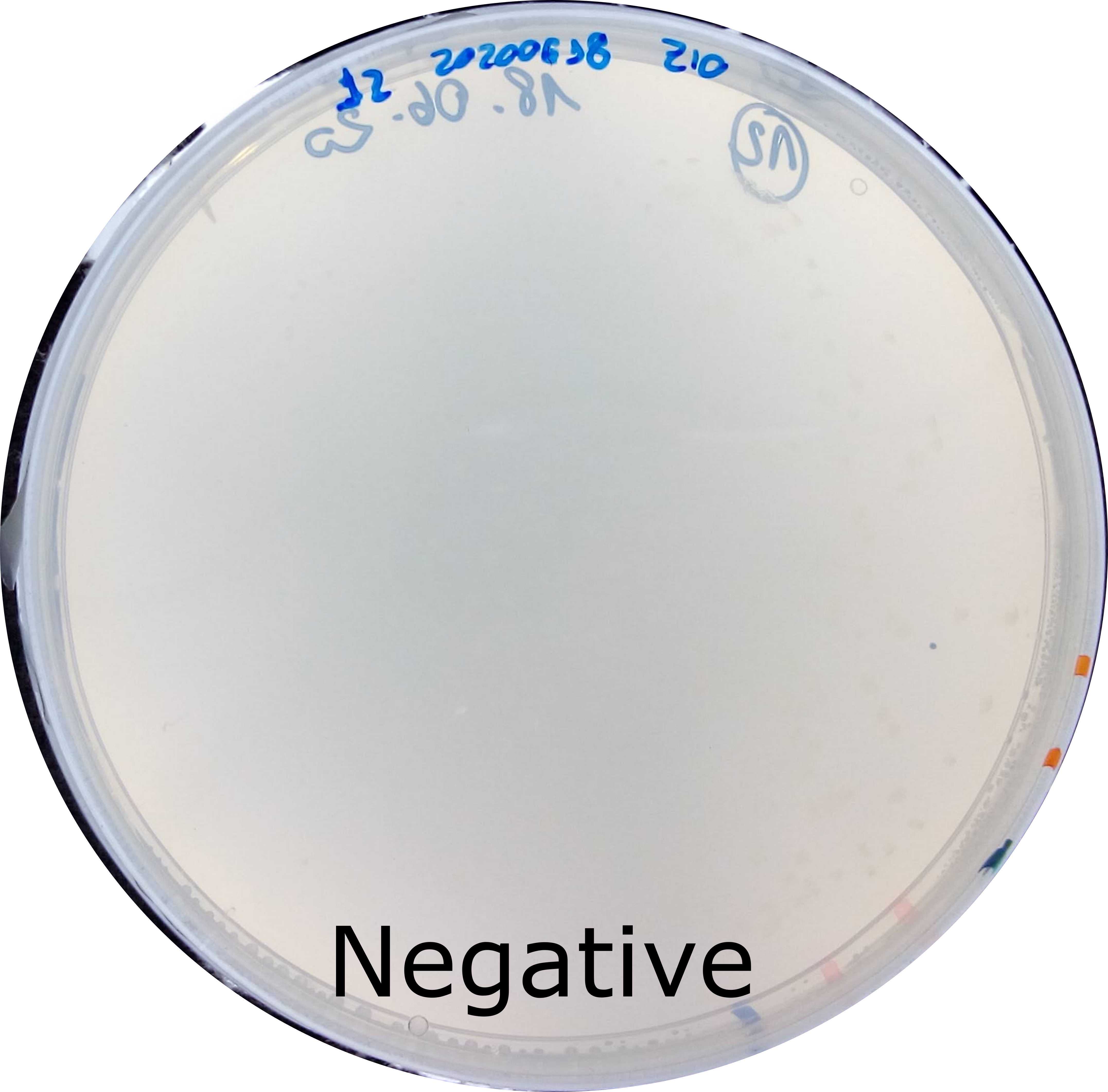
Add cutted vector to the bottom of the electroporation cuvette. Typically from 250ng to 1000ng Add 250µL (at approximatelly 3x 10^8 cells/mL) to each cuvette. Pippet up and down on DNA sample. Flick cuvette to mix DNA and cells. Shake cells to the bottom of the cuvette. Also add no DNA control (Elution buffer or water).
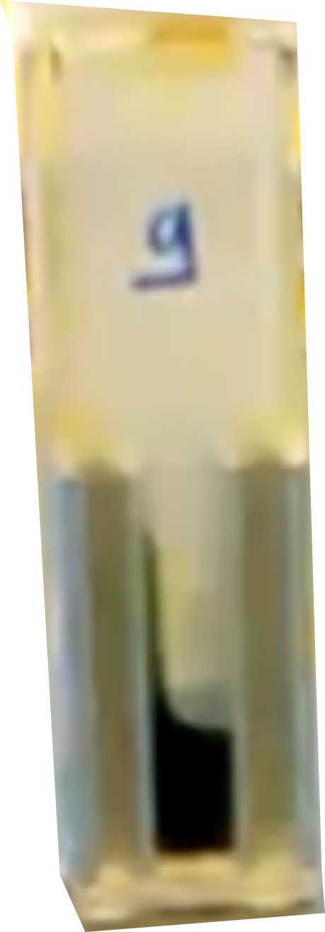
- Incubate cells with DNA
On icefor0h 10m 0s - Wipe cuvette (to remove condensated water) and electroporate (Table Electroporation).
- Let it recover for
0h 10m 0son the cuvette - Add cells to
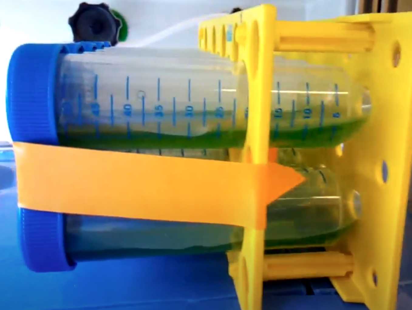
10mLinside sterile 50mL centrifuge tubes. Gently transfer cells from cuvette to TAP/40 mM sucrose. Rinse cubette with TAP/40 mM sucrose to transfer any remaining cells. Incubate atRoom temperatureon rocker or shaker at 50 rpm ambient light.
- Pellet cells by centrifuging for
2000x g,25°C - Aseptically poor off supernatant. Add
300µLto pelet. Gently re-suspend cells and pipette onto 2 plates with appropriate antibiotics. ie.200µLper plate, and let it dry a,septically without plate cover. - Spread cells evenly over the plate with a innoculation loop. Avoid spreading to the borders.
- Use parafilm to block evaporation and place plates under constant light (60 µmols de photons/m2s),
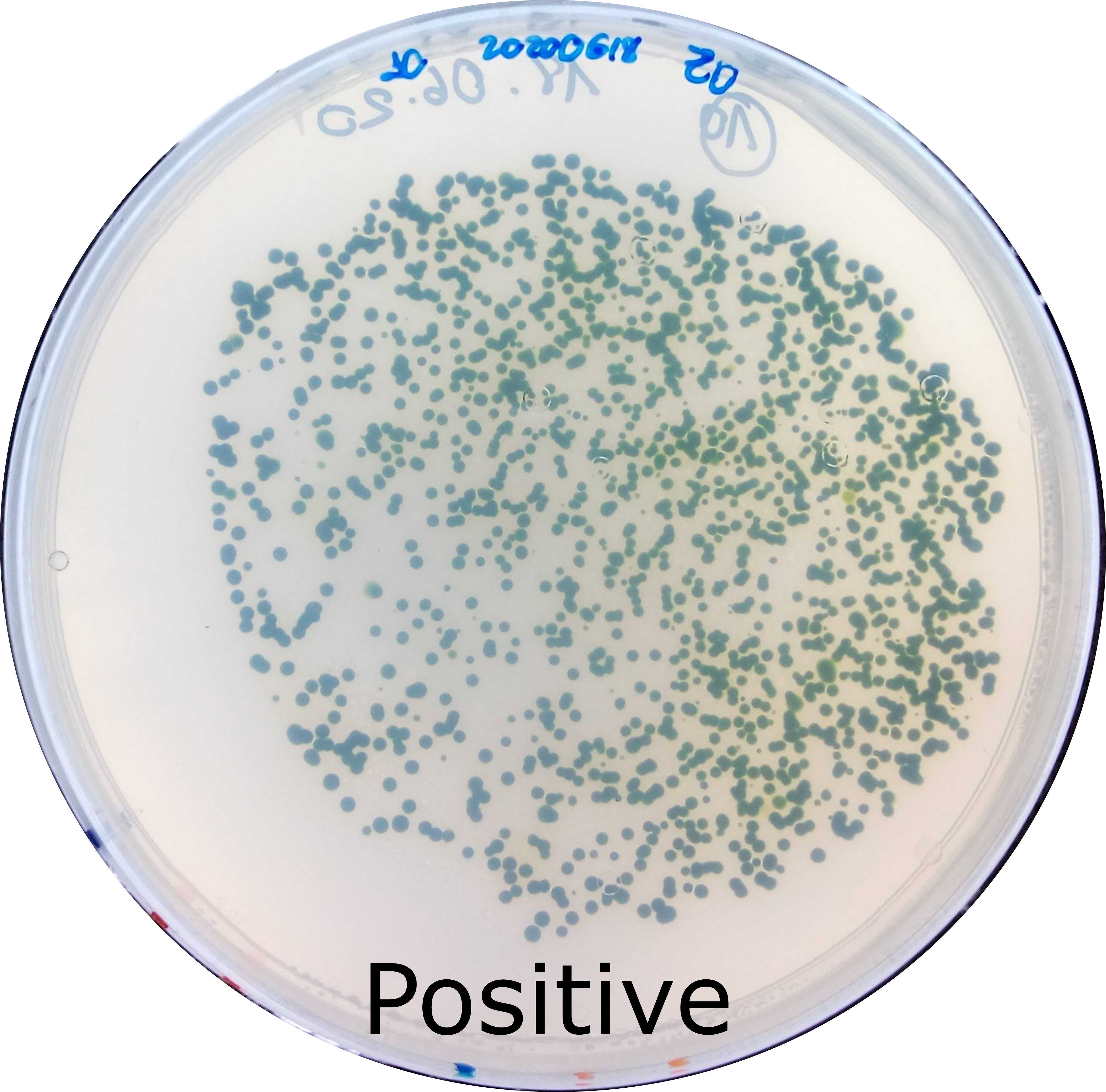
25°C. Colonies should be visible in 5-7 days.
Table Electroporation - Settings
| A | B |
|---|---|
| Voltage | 800 V |
| Time Constant | 20 ms |
| Cuvette gap | 4 mm |
Equipment
| Value | Label |
|---|---|
| Gene Pulser Xcell Electroporation Systems | NAME |
| Electroporator | TYPE |
| Biorad | BRAND |
| 1652660 | SKU |
Typical output after electroporation
| A | B | C | D |
|---|---|---|---|
| Time constant (ms) | Voltage (V) | Capacitance (uF) | Resistance ( Ohms) |
| 20.1 | 788 | 50 | 650 |
| 20.4 | 789 | 50 | 600 |
| 19.8 | 789 | 50 | 550 |
