Bovine necropsy
Laetitia Dorso
Abstract
A necropsy is the final medical diagnostic procedure performed on a deceased animal to determine the cause of death.
The aim of a necropsy is to investigate and define as precisely as possible the cause of death of the animal, or to identify and confirm the suspected pathological process.
It is a medical examination. It consists of several stages, and must be carried out in premises dedicated to this activity, in accordance with the regulations.
A summary report must be drawn up and signed by the pathologist to record the observations made. The samples and analyses, if needed, must be taken as soon as possible after the body'arrival at the laboratory.
Steps
Anamnesis
Breeding:
Rearing method, number and age of animals
Feed
Breeding health plan, vaccines, deworming, others (nature, dates)
Morbidity, mortality (diagnosis, number and %)
Animal:
Date of introduction in the herd
Symptoms (description, chronology, duration)
Conditions, date and time of death
Treatments (nature, date)
Complementary examinations (nature, dates, results)
Necropsy technique for an adult cattle
The animal is suspended by one hind leg.
Check the animal's identity (Conforming buckle)
Assessment of the condition of the animal
- Observation of the skin (erosion, etc.), overall silhouette (swelling, oedema, amyotrophy, etc.)
- Observation of mucous membranes (eyes, mouth) colour changes (congestion, pallor, cyanosis, petechiae)
- Observation of the eyes and oral cavity
- Observation of the hindquarters and rectal and vaginal mucosa looking for signs of diarrhoea, congestion and petechiae. A T-shaped incision is made: the two horizontal branches of the T follow an axis from the caudal linea alba to the lumbar spine, and the vertical branch of the T runs from the pubic symphysis to the xiphoid process along the linea alba.
The vertical incision involves the skin, abdominal muscles and peritoneum.
- Observation of the peritoneal cavity (presence of exudate, blood) and parietal peritoneum.
- Identification of the body condition score based on observation of pelvic adipose tissue.
- Section at distal rectum level. Downward reclination of the digestive mass (using gravity) after section of all mesos along the spine.
Analysis of abdominal viscera
-
Isolate the spleen, liver and kidneys: make multiple sections in the liver parenchyma and a longitudinal section of the kidneys after decapsulation.
-
Separation of the foregut from the intestinal mass.
-
Opening of the foregut successively according to their greatest curvature (rumen, reticulum, omasum, abomasum).
-
Observe the contents of the foregut: qualitative and quantitative aspects.
-
Observe the mucosae. Lay out the intestinal mass so as to identify the main segments (on one side duodenum, jejunum, ileum, ileocaecal valve, caecum, mesenteric lymph nodes, on the other side spiral colon, rectum).
-
Open each segment (duodenum, proximal jejunum, medial jejunum, distal jejunum, ileum, caecum, spiral colon and rectum) to a length of approximately 10 cm and observe the contents (qualitative/quantitative) and the appearance of the mucosa.
-
Open the uterus: observe the contents and mucosa.
-
Open the bladder: observe the contents and mucosa. Analysis of the thoracic viscera:
-
Cut the diaphragm where it attaches to the ribs and check for the presence of a pleural vacuum.
-
Observe the pleural cavity (presence of exudate, blood) and the parietal pleura.
-
Cut the oesophagus and trachea cranially to remove the heart/lung block.
-
Separate the heart from the lungs.
-
Open the oesophagus longitudinally: analyse any contents and the oesophageal mucosa
-
Open the trachea to the tracheobronchial junction: analyse any contents and the tracheal mucosa.
-
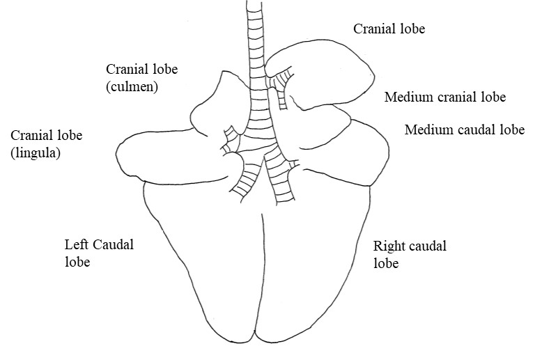
Palpate the lung parenchyma carefully (cranially first, then caudally), looking for any change in consistency.
-
Make a section in each of the lobes and observe: the airways, the vessels, the alveolar parenchyma.
-
Cutting the heart: the pericardium is incised and the heart is removed. A first apical section is made about 1/3 of the apex.
-
The ventricular walls are examined and their thickness assessed. The right heart is placed on the left and the left heart on the right.
-
A second incision is made in the left ventricle, following the septum and opening the left ventricle and aorta.
-
A third incision, parallel to the second, is made through the left ventricle and left atrium.
-
A fourth incision is made in the right ventricle, following the septum and opening the right ventricle and pulmonary artery.
-
A fifth incision, parallel to the fourth, is made through the right ventricle and right atrium.
-
Observation of heart valves and myocardium
-
Observation of lymph nodes in the carcase
-
Observation of joints and feet
Record each observation in the following sections, using the tables provided.
Identification
| A | B | C | D |
|---|---|---|---|
| Ear tag | Sex | Age | Breed |
Necropsy findings
| A | B | C | D |
|---|---|---|---|
| Body Condition Score / 5 : | □ Correct | □ Amyotrophy | □ Leanness |
| A | B | C | D |
|---|---|---|---|
| Status of preservation | □ Correct | □ Poor | □ Autolysed |
| A | B | C | D | E | F |
|---|---|---|---|---|---|
| Exterior appearance | □ No significant lesion (NSL) | □ Diarrhoea spots | □ Limb swelling | □ Scabs | □ Other lesions |
| A | B | C |
|---|---|---|
| Abdominal cavity | □ NSL | |
| □ Effusion | □ Hemorrhagic | |
| □ Sero-hemorrhagic | ||
| □ Fibrinous | ||
| □ Suppurated | ||
| □ Adhesions | □ Suppurated | |
| □ Fibrosis |
| A | B | C | D | E |
|---|---|---|---|---|
| Oral cavity | □ NSL | □ Ulcerations | □ Other | |
| Teeth | □ NSL | □ Abnormal coloration | □ Abnormal abrasion | □ Other |
| Tongue | □ NSL | □ Ulcerations | □ Other | |
| Oesophagus | □ NSL | □ Ulcerations | □ Obstructions | □ Other |
| A | B | C | D | E | F |
|---|---|---|---|---|---|
| Rumen | Content | □ NSL | □ Dry | □ Liquid | □ Other |
| Mucosa | □ NSL | □ Ulcers | □ Melanosis | □ Other | |
| Reticulum | Content | □ NSL | □ Dry | □ Liquid | □ Other |
| Mucosa | □ NSL | □ Ulcers | □ Melanosis | □ Other | |
| Omasum | Content | □ NSL | □ Dry | □ Liquid | □ Other |
| Mucosa | □ NSL | □ Ulcers | □ Melanosis | □ Other | |
| Abomasum | Content | □ NSL | □ Dry | □ Liquid | □ Hemorrhagic |
| Mucosa | □ NSL | □ Ulcers | □ Hemorrhagic | □ Fundus | |
| □ Chronic | □ Pylorus |
| A | B | C | D | E | F |
|---|---|---|---|---|---|
| Duodenum | |||||
| Content | □ NSL | □ Hemorrhagic | □ Fibrinous | □ Parasites | □ Other |
| Mucosa | □ NSL | □ Congestive | □ Fibrin | □ Necrosis | □ Other |
| Jejunum | |||||
| Content | □ NSL | □ Hemorrhagic | □ Fibrinous | ||
| Mucosa | □ NSL | □ Congestive | □ Fibrin | □ Necrosis | □ Thickening |
| □ Parasites | □ Other | ||||
| Ileum | |||||
| Content | □ NSL | □ Hemorrhagic | □ Fibrinous | □ Other | |
| Mucosa | □ NSL | □ Congestive | □ Fibrin | □ Necrosis | □ Thickening |
| □ Parasites | □ Other | ||||
| Colon | |||||
| Content | □ NSL | □ Hemorrhagic | □ Fibrinous | □ Parasites | □ Other |
| Rectum | |||||
| Content | □ NSL | □ Hemorrhagic | □ Fibrinous | □ Parasites | □ Other |
| Mucosa | □ NSL | □ Congestive | □ Fibrin | □ Necrosis | □ Other |
| Mesenteric lymph nodes | □ NSL | □ Increased size | □ Necrosis | □ Suppuration |
| A | B | C | D | E |
|---|---|---|---|---|
| Liver | Size | □ NSL | □ Increased | □ Reduced |
| Shape | □ NSL | □ Rounded | ||
| Color | □ Red | □ Beige | □ Orange | |
| Consistency | □ NSL | □ Increased | □ Reduced | |
| □ Abscess | □ Necrosis | |||
| Gall bladder | □ NSL | □ Repletion | □ Emptiness |
| A | B | C |
|---|---|---|
| Pleural cavity | □ NSL | |
| □ Effusion | □ Hemorrhagic | |
| □ Sero-hemorrhagic | ||
| □ Fibrinous | ||
| □ Suppurated | ||
| □ Adherences | □ Fibrinous | |
| □ Fibrosis |
| A | B | C | D | E | F |
|---|---|---|---|---|---|
| Nasal cavities | □ NSL | □ Congestion | □ Fibrin | □ Necrosis | □ Suppuration |
| Trachea | □ NSL | □ Congestion | □ Fibrin | □ Ulcer | □ Foam |
| Larynx | □ NSL | □ Congestion | □ Fibrin | □ Ulcer | □ Foam |
| Lungs | Size | □ NSL | □ Increased | □ Reduced | |
| Shape | □ NSL | □ Rounded | |||
| Color | □ Pink | □ White | |||
| Lesion characteristics (if lung involvement) | |||||
| Distribution | □ Focal | □ Multifocal | □ Extensive | □ Disseminated | |
| Affected percentage | □ 0-25% | □ 25-50% | □ 50-75% | □ 75-100% | |
| Demarcation | □ Clear | □ Blurred | |||
| Exudate | □ NSL | □ Foam | □ Mucus | □ Suppurated | |
| Tracheobronchial lymph nodes | □ NSL | □ Increased size | □ Necrosis | □ Suppurated |
| A | B | C | D | E |
|---|---|---|---|---|
| Heart | Pericardium | □ NSL | □ Hemorrhagic | |
| □ Sero-hemorrhagic | ||||
| □ Fibrinous | ||||
| □ Suppurated | ||||
| Myocardium | □ NSL | □ Hypertrophy | □ Necrosis | |
| Valve | □ NSL | □ Endocarditis | ||
| Abnormalities | □ Ventricular septal defect | □ Atrial septal defect | □ Patent ductus arteriosus |
| A | B | C | D | E |
|---|---|---|---|---|
| Kidney | Size | □ NSL | □ Increased | □ Dicreased |
| Shape | □ NSL | □ Modified | ||
| Color | □ Red | □ Beige | □ Other | |
| Consistency | □ NSL | □ Increased | □ Dicreased | |
| □ Tumor infiltration | □ Necrosis | □ Other |
Tumour infiltration
| A | B | C | D | E | F |
|---|---|---|---|---|---|
| Joint | Joint cavity | □ NSL | □ Hemorrhage | □ Fibrin | □ Suppuration |
| Joint cartilage | □ NSL | □ Erosion | □ Necrosis | ||
| Bones | □ NSL | □ Osteomyelitis | |||
| Muscles | □ NSL | □ Hemorrhage | □ Necrosis | □ Degeneration | |
| A | B | C | D | E |
|---|---|---|---|---|
| Udder | Color | □ Beige | □ Red | □ Purplish |
| Size | □ NSL | □ Increased | □ Dicreased | |
| Consistency | □ NSL | □ Increased | □ Dicreased | |
| □ Abscess | □ Milk modification | □ Necrosis |
| A | B | C | D | E |
|---|---|---|---|---|
| Genital system | ||||
| Ovary / testicle | □ NSL | □ Other | ||
| Penis | □ NSL | □ Other | ||
| Uterus | □ NSL | □ Pregnant | □ Exudate | □ Hemorrhagic |
| □ Suppurated | ||||
| □ Necrotic |
| A | B | C | D | E |
|---|---|---|---|---|
| Brain | □ NSL | □ Hemorrhage | □ Necrosis | □ Abscess |
| Meninges | □ NSL | □ Congestion | □ Fibrin | □ Suppuration |
Assesment of lesions
Assessment of lesions
Significant lesions
Non significant lesions
Conclusion
