Automated Immunohistochemistry Staining
Hemanth Ramesh Nelvagal, Toby J Curless, Zane Jaunmuktane
ASAPCRN
Immunohistochemistry
Brain sections
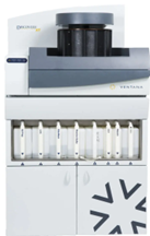
Ventana Discovery® XT Immunostainer
Antibody diluent solution (Ventana)
Abstract
The protocols describes the steps for automated immunohistochemistry staining using the Ventana Discovery® XT
Immunostainer.
Steps
Preparation
Heat tissue dry tissue sections for 1h 0m 0s at 60°C
Generate tissue sections using standard microtome sectioning protocols.
Prepare primary and secondary antibody solutions as per manufacturer's protocol - see below for example for AT8.
Primary AT8 antibody (Thermo Fisher Scientific, MN1020) diluted at 1:100 in antibody diluent solution
(Ventana). Secondary anti-mouse antibody (Abcam) diluted at 1:100 in antibody diluent solution (Ventana).
Perform immunohistochemical staining using the Ventana Discovery® XT Immunostainer – following manufacturers guidelines.
Print barcoded slide labels corresponding to the correct protocol on the Ventana machine and stick them to the top of each slide following the slide heating.
Processing Slides Using the Ventana Machine
Place slides into the Ventana machine and ensure all bulk reagents are sufficiently filled
Begin the staining protocol.
Upon completion of the staining, activate the counterstaining step.
Following counterstaining, remove the slides and wash in soapy water 5x.
Leave slides in running water for 0h 5m 0s mins.
To deparaffinise sections, place slides in 100 % ethanol for 0h 5m 0s mins, remove and place in separate 100 % ethanol for 0h 5m 0s mins. Remove from ethanol and place in xylene for 0h 5m 0smins, remove and place in separate xylene for0h 5m 0smins
For cover-slipping of slides following deparaffinisation – use the Leica CV5030 automated slide cover-slipping machine.
Stained slides digitised on a NanoZoomer S360 scanner (Hamamatsu) – Brightfield scan profile at x40 magnification.
Upload digitised slides to NZ Connect (1.0.36 (IVD)) slide viewing platform.


